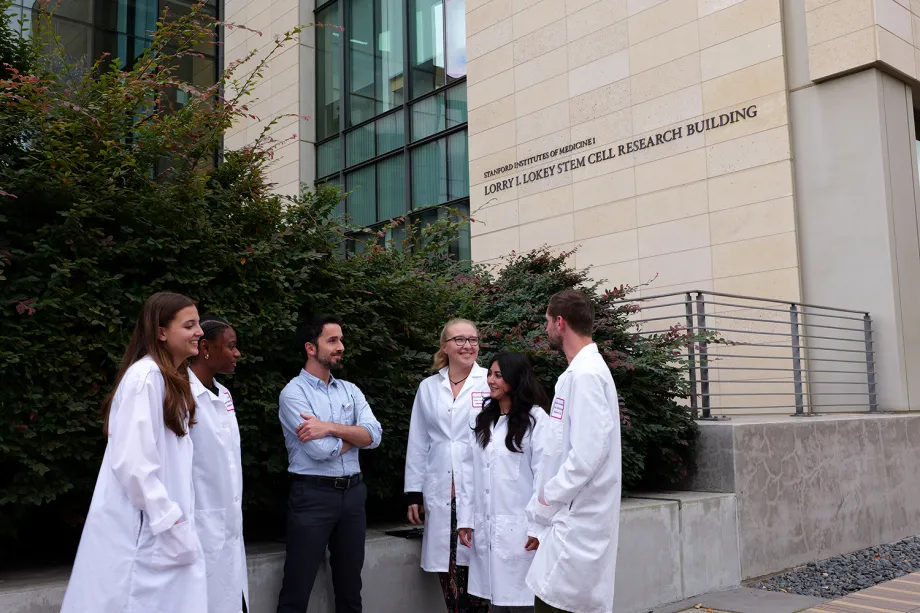
Matteo Molè (center), assistant professor of obstetrics and gynecology, and stem cell PhD student Nicole Horsley (to his right, wearing glasses) are part of a team that's developed a novel 3D uterus model. | Courtesy Stanford Medicine
Stanford Report News - May 27th, 2026 - Cassie Myers
Science has advanced enormously over centuries, but there are still countless mysteries. One such “black box,” as Matteo Molè, assistant professor of obstetrics and gynecology, describes it, arises during the early stages of pregnancy, when the embryo attaches or “implants” onto maternal tissue in the endometrium.
It’s a process that occurs inside the uterus where no scientist can see it, a process that stymies millions of couples suffering from infertility and undergoing IVF. So seeing inside this black box could be the key to unlocking fertility treatments, helping create therapies for uterine and pregnancy-related diseases, or simply understanding how fertility and successful pregnancies occur.
Molè, a member of the Stanford Institute for Cell Biology and Regenerative Medicine (ISCBRM), found one potential solution: create a 3D model of the uterine wall in a lab setting and then observe how embryos attach (or don’t attach). This work, published recently in Cell, could be a game changer.
As Molè explains, an estimated 30-60% of embryos are lost at this implantation stage, which is the primary factor behind the success rates for IVF (in-vitro fertilization, which creates a viable embryo in the lab and then implants it in the uterus). In fact, success rates for IVF are currently below 40-50% even in the best cases.
“It’s pretty shocking that this critical process is so inefficient,” Molè says. “So many couples undergo multiple cycles of IVF, often unsuccessfully. And we don’t know what’s going on because nobody has ever managed to study it at this stage.”
But if you recreate this process in a petri dish, you can. And this is precisely what Molè did: first at Cambridge, where he began his postdoctoral work, and then at Stanford, where he came to the ISCBRM as an assistant professor and started his own lab.
Building the black box
So – how do you go about building a lab model of this previously hidden process? First, you build a mini uterus from tissue biopsied from consenting patients. Next, you take fertilized embryos donated from couples undergoing IVF and you transfer them into the modelled uterine tissue – and then you can observe the process outside of the uterus in real time.
“It’s beautiful to see the initial contact between embryo and maternal tissue in this kind of detail,” Molè says. “If you think about it, there are two different organisms interacting here, the embryo and the mother, who need to communicate with each other to establish a successful pregnancy. And that’s the big question: how these two interact on a molecular level, why most of the embryos fail to implant, and some succeed, and how the two entities communicate. This is what we’re really trying to understand in the lab.”
What’s next
The model’s construction was the first hurdle – but now that it’s up and running, it can be used for many different types of experiments.
For one thing, it will allow researchers to study the formation of the human placenta – the tissue that promotes the flow of oxygen and nutrients from the mother to the growing fetus. And Molè’s next project will engineer the maternal vasculature (specifically arteries and vessels from the mother’s uterine tissue that interact with the placenta from the embryo) as well.
“We’re approaching this from many different points of view,” he explains, “from transcriptomics of gene expression analysis to proteomics to studying the secretions and primary contacts between embryo and maternal uterine tissue.”
But even that’s not all: the Molè lab is also considering the role placental and uterine tumors and genetics may play in the process, and whether and how all these factors can “impede or augment” embryo implantation.
‘A big gamble’
The model may be the jumping-off point for future research, but it’s also a culmination of years of Molè’s work both at Cambridge and Stanford. “It was a big gamble,” he says. “We didn’t know if we would be able to successfully recreate the process of implantation in vitro.”
It took years to successfully build the model, and writing and revising the initial publication took months more. Molè worked with several researchers between Cambridge and Valencia and found one crucial student in his own Stanford lab: Nicole Horsley, a stem cell PhD candidate. She joined the process when she started at the Molé lab in 2024, and led key experiments during the paper’s revision phase that characterized and validated the model, helping establish it as a viable research tool for implantation.
“Nicole has been instrumental to the project, considering how much work she did in such a short time,” Molè says. So instrumental, in fact, that Molè named her co-first author of the publication.
Horsley joined Molè’s lab midway through her PhD after realizing that her original lab was not the right scientific fit. She first worked in Molè’s lab during a short rotation, a process where PhD students spend time in different labs before choosing a doctoral home. During that rotation, she “fell absolutely in love with early development, embryos, and blastoids,” and she has been working with him ever since.
“A big piece of the paper was my work comparing what happens when embryos are grown on their own versus when they interact with maternal tissue,” she explains. In other words, she was studying how early embryos develop with and without support from the cells that normally line the uterus. “I did this really big experiment first with blastoids, stem cell-based embryo models, and then with actual human embryos, to show that without that uterine environment, we see much less development of the cells which are vital to building the placenta.”
She’s as excited as he is by the doors this research may open. “Right now, many of the tools available to scientists offer only a fixed end point: they can show what implantation looks like after a particular moment has passed, but not the dynamic sequence of events that got it there,” she explains. Her personal next step will be trying to capture “live imaging” of implantation, essentially a movie of the full implantation process, so researchers can watch in real time as it unfolds.
“Seeing the dynamics happen in real time is something I feel really passionate about,” she says. “There’s so much to be learned about early embryo development from this model. It’s really, really exciting. [For me, the science behind how] a small distribution of cells becomes this super complex organism or organ is really cool.”
Micro to macro
The work can be microscopic, but Horsley also sees the bigger picture. “I feel like women’s health is in general understudied,” she adds, “especially diseases of the endometrium and fertility more broadly. IVF is expensive, often unsuccessful, and inaccessible to many families. I’d love to use this model to help develop noninvasive or less expensive fertility treatments, and to learn more about diseases of pregnancy, like preeclampsia. This research has this incredible potential to make a real difference for people trying to build families, while also opening the door to fascinating new science.”
The real-world impact is not just in individual families, either – as Molè points out, fertility issues have economic and political implications as well, with declining birthrates and aging populations worldwide. IVF has already been responsible for millions of births, and further leaps in fertility treatments could help countless people.
“If we can translate this knowledge into therapeutic interventions and improve the efficiency of that process, patients won’t have to go through multiple failed cycles of IVF,” he says. “This will save patients a lot of money but also reduce the profound emotional and psychological toll of failure, giving them that possibility to get it right from the very first embryo transfer. So for me, it’s a very exciting time.”
“[This research] has implications for everyone, including those in the LGBT community that I’m very close to,” he adds. “I’m hoping it’s going to have positive ramifications for everyone – for any patients who want to build a family.”