Photo by DrimaFilm, Shutterstock.
Stanford Medicine Scope - April 13th, 2017 - by Krista Conger
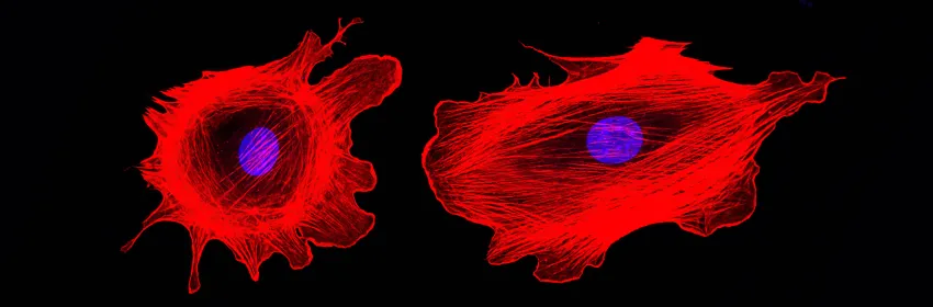
Odds are, you haven’t spent a lot of time thinking about the cytoskeleton that props up your cells and gives them their three-dimensional structure. But maybe you should. Like Tinkertoys of old (am I dating myself here?), bits of a protein called actin assemble into microfilaments necessary for many critical cellular processes. Without these actin filaments, many of your cells would likely collapse into little functionless blobs of goo.
Now neurobiologist Brad Zuchero, PhD, in collaboration with colleagues from the laboratories of neurobiologist Ben Barres, MD, PhD, and Casper Hoogenraad, PhD, from Utrecht University in the Netherlands, have come up with a tool that will allow researchers to study the cytoskeleton with much greater precision than has previously been possible. They published their results this week in Nature Methods.
As Zuchero described to me in an email:
Basically, we’ve created a genetic toolkit that enables researchers to perturb any cell’s cytoskeleton — the cell skeleton that gives cells shape, lets them move, and, when misregulated in cancer, makes tumor cells invade nearby tissues.
Historically, scientists interested in understanding the role of the cytoskeleton in cell motility focused on single cell types like immune cells growing in a laboratory dish. Drugs that disrupted the cytoskeleton didn’t have to be cell-type specific for the researcher to gain valuable insights because there was often only one type of cell in the dish. This approach is inadequate, however, if researchers wanted to study whole organisms or multiple cell types.
Zuchero and his colleagues came up with a new genetic approach they termed DeActs, which borrows a strategy from a potent pathogen. As he explained:
In order to engineer tools that specifically perturb the actin cytoskeleton, we turned to nature. Salmonella and other pathogenic bacteria have evolved potent toxins that they use to infect epithelial or immune cells when we eat or drink contaminated food or water. We repurposed one of these toxins — an enzyme that triggers actin cytoskeleton disassembly in infected cells — as a universal tool for perturbing actin.
This genetic approach allows the researchers to specifically trigger the disassembly of the actin cytoskeleton in individual cell types — pulling apart the Tinkertoy structure, if you will — by selectively inducing them to express the actin-disrupting proteins. They can then study the effect of disrupting the cytoskeleton specifically in developing neural cells in an embryonic mouse brain, for example. But the approach is likely to be broadly applicable to many types of research. In particular, Zuchero is interested in learning about the relationship between the cytoskeleton and the myelination (or coating) of neurons necessary for the conduction of nerve impulses.
As Zuchero explained:
Previously we had discovered that, completely unexpectedly, the actin cytoskeleton is dismantled at the start of myelin wrapping. Our main goal now is to use this DeActs tool to understand how actin disassembly promotes myelin wrapping, and whether and how this might be important in multiple sclerosis, in which myelin attacked by the immune system fails to re-wrap around the neuron. We hope that by understanding the mechanisms of myelin formation we will be able to promote its regenerations in diseases like multiple sclerosis. Other researchers may choose to use DeActs to investigate how cancer cells metastasize or how neurons wire themselves properly during development.