Interdisciplinary Initiatives Program Round 4 – 2008
Daniel Bernstein, Pediatrics
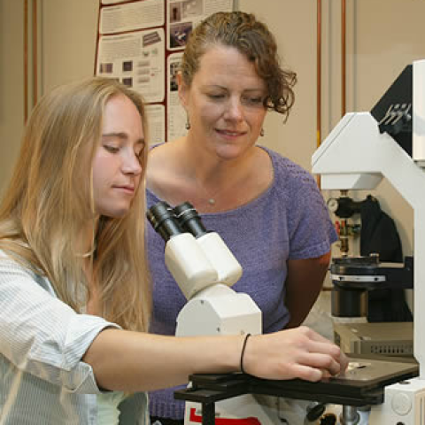
Beth Pruitt, Mechanical Engineering
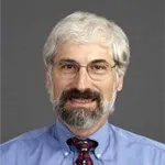
Philip Yang, Cardiovascular Medicine
Cardiovascular disease is the leading cause of death in the United States. Of particular importance in this epidemic is the reality that cardiac muscle cells cannot self-renew; thus, tissue damaged during a heart attack does not regenerate. The remaining cells do, however, remodel the tissue, causing a decrease in function and ultimately resulting in heart failure. The exact response which results in heart failure, however, is poorly understood. While we can observe morphological and physiological changes in the heart after a heart attack, we lack detailed information about mechanical changes on the cellular level.
Cardiac muscle cells are known as cardiomyocytes (CMs). It is well known that stem cells can be differentiated to CMs. Stem cell therapy is a promising methodology for regenerating the CMs. However, these cells will be a viable replacement therapy only if their biomechanical properties recapitulate those of healthy native CMs. Several genetically altered mouse models have been developed to mimic the pathophysiology of cardiovascular disease in humans. Although genetic alteration is possible in any species, the mouse has been the most often used because of their well-defined genetic backgrounds, ease of genetic manipulation, and relatively low costs. Comparative study of biomechanical properties among isolated CMs from healthy and diseased mouse, and mouse embryonic stem cell derived CMs will provide insightful information about cardiovacular disease. In order to measure mechanical properties at a cellular level, an experimental platform capable of measuring nanoNewton force is required. In collaboration between Mechanical Engineering and School of Medicine at Stanford, the experimental platform based on a micrometer-sized compliant post array microfabricated by Dr. Pruitt’s group in Mechanical Engineering enable us to mechanically evaluate CMs isolated from the adult mouse. Dr. Bernstein’s group in Pediatric Cardiology is able to induce several different types of heart failure in mice and isolates CMs from healthy and diseased mice to investigate the cellular mechanisms of chronic heart failure. By enhancing our knowledge of mechanical characteristics of normal and diseased adult CMs, we will then be able to use mouse embryonic stem cell derived CMs prepared by Dr. Yang’s group in Cardiovascular Medicine to characterize the therapeutic effectiveness of stem cells for treating heart muscle disease.
These studies will provide a better understanding of how cardiac function can be rescued at the cellular level, how to improve conditions for stem cell differentiation, and will provide a high throughput test bed for new pharmacologic, small peptide, and gene therapies. These techniques will be essential for the future assessment of human pluripotent-cell derived CMs.