Welcome to the biweekly electronic newsletter from Stanford Bio-X for members of the Bio-X Corporate Forum. Please contact us if you would like to be added or removed from this distribution list, or if you have any questions about Stanford Bio-X or Stanford University.
Highlights
** On October 9, 2013, Bio-X celebrated the 10th Anniversary of the James H. Clark Center, the hub of Bio-X. Check out CLARK CENTER @ 10X on the SPLASH PAGE as well as the Bio-X Timeline over the last 15 years!!
** Check out the article by Stanford President John Hennessy in the Nov/Dec 2013 issue of the Stanford Magazine on Bio-X and the Clark Center, "A Cauldron of Innovation".
Matthew Scott to become president of Carnegie Institution for Science
 Previous Bio-X Director to become president of Carnegie Institution for Science
Matthew Scott, PhD, professor of developmental biology, of genetics and of bioengineering, will become the president of the Washington, D.C.-based Carnegie Institution for Science in September. Scott, who is also the Howard H. and Jessie T. Watkins University Professor, has been at Stanford since 1990. "Matt has been an invaluable leader, scientist and colleague during his years at Stanford," said Lloyd Minor, MD, dean of the School of Medicine. "His early studies of the regulators of early embryonic patterning set the stage for many groundbreaking studies in the field of developmental biology, and his experience as the chair of Bio-X immersed him in the type of interdisciplinary research for which the Carnegie Institution is known. We are sorry to see him go, but we are excited for him and his future in this new phase of his career. He is eminently qualified for the position." Click here to read the entire article!
Seed Grants
**UPDATE: Bio-X has closed the 7th RFP for its IIP Seed Grants, and review of the 141 Letters of Intent is underway!
 SEED GRANTS FOR SUCCESS - Stanford Bio-X Interdisciplinary Initiatives Program (IIP)
The Bio-X Interdisciplinary Initiatives Program represents a key Stanford Initiative to address challenges in human health. The IIP awards approximately $3 million every other year in the form of two-year grants averaging about $150,000 each. From its inception in 2000 through the fifth round in 2010, the program has provided critical early-stage funding to 114 different interdisciplinary projects, involving collaborations from over 300 faculty members, and creating over 450 teams from five different Stanford schools. From just the first 5 rounds, the IIP awards have resulted in a 10-fold-plus return on investment, as well as hundreds of publications, dozens of patents filed, and most importantly, the acceleration of scientific discovery and innovation.
In 2012, Stanford Bio-X selected 23 new seed grant projects as the winners of the 6th round. Please go here to view the list of awardees, along with the titles of their projects and the abstracts of the research. Competition was intense as the awardees were chosen from 118 Letters of Intent (LOIs). Selection criteria included innovation, high-reward, and interdisciplinary collaboration. (To view the 114 other IIP projects that have been funded from the first 5 rounds, please click here.) In addition, SANOFI has also funded 4 new Bio-X IIP Seed Grant projects from round 6!
We are cultivating and are highly successful in building meaningful collaborations with numerous corporate colleagues. New collaborations through our seed grant projects are highly encouraged. To learn about how to get involved, please contact Dr. Hanwei Li or Dr. Heideh Fattaey.
IIP Seed Grants-Related Events
** On Monday, March 3, 2014, Bio-X had a Poster Session, featuring 105 different posters from research by all scientists within the Stanford Bio-X community. Over 250 people attended the session, which allowed for an excellent venue to discuss science and research with colleagues from both academia and industry.
** On Monday, August 26, 2013, Bio-X had its second annual IIP Symposium of the year at the Clark Center, which highlights projects that exemplify the Stanford Bio-X mission of crossing boundaries to bring about interdisciplinary research and solutions in the field of life bioscience. The symposium was a huge success with over 300 people attending this event, which included 8 oral presentations and 136 poster presentations. Recorded talks from the symposium will be uploaded soon. If you'd like to view the talks for previous symposia through the years, please click here.
Fellowships
**UPDATE: Bio-X has closed the RFP for its 11th annual Bio-X Graduate Student Fellowships, and received 100+ submissions.
 BIO-X FELLOWSHIPS
Every year, graduate students and postdoctoral scholars of Bio-X affiliated faculty are highly encouraged to apply for the Bio-X Fellowships, which are awarded to research projects that are interdisciplinary and utilize the technologies of different fields to solve different biological questions. Students are encouraged to work collaboratively with professors of different departments, thus creating cross-disciplinary relationships among the different Stanford schools. Our fellows have conducted exciting research, resulting in publications in high-impact journals and have been offered excellent positions in industry and academia. To date, Stanford Bio-X has a total of 152 Fellows.
On June 26, 2013, Bio-X held its annual Bio-X Fellows Symposium, where there were four 15-minute oral presentations followed by one-minute spiels from current fellows. The 25 newest fellows selected this year were also announced, and about 100 attendees came to the symposium. Please click on the "Bio-X Fellows Symposium" link above for the agenda and titles of the talks, and on the icon of the brochure above for the updated and latest Bio-X Fellowships brochure.
To view the numerous projects that have been awarded over the years, please click here.
**UPDATE: Bio-X has closed the RFP for its 9th annual Bio-X USRP, and received 154 applications.
 BIO-X UNDERGRADUATE SUMMER RESEARCH PROGRAM
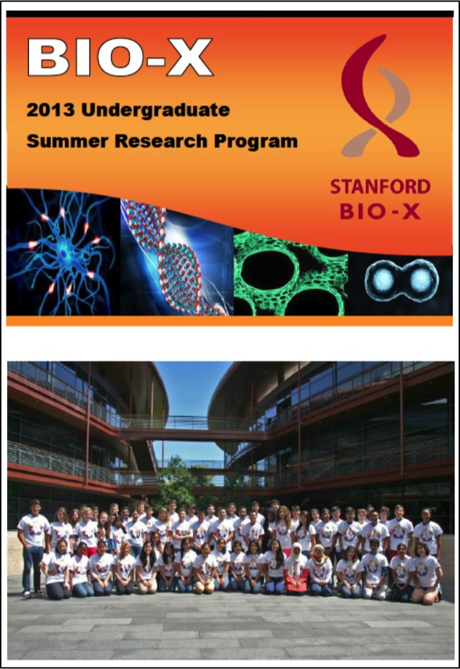
The Bio-X Undergraduate Summer Research Program supports undergraduate research training through an award designed to support interdisciplinary undergraduate summer research projects. The program is an invaluable opportunity for students to conduct hands-on research, learn how to carry out experiments in the laboratory, and develop the skills to read and analyze scientific literature.
This program is eligible to Stanford students who want to work in the labs of Bio-X affiliated faculty. To date, 241 students have been awarded the opportunity to participate in the Bio-X Undergraduate Summer Research Program. This summer is Stanford Bio-X's 8th round of USRP.
Participating undergraduates are also required to present poster presentations on the research that they've conducted during the program. Please click here for title lists of past posters that our undergraduates have presented.
Many fruitful collaborations and relationships have been established with industry through fellowships. Please contact Dr. Hanwei Li or Dr. Heideh Fattaey if you'd like to learn more about how to get involved with these fellowship programs.
News
 Innovative Training for Biomedical Technology
Bio-X Affiliated Faculty and Biodesign Director Paul Yock
One common approach to academic entrepreneurship in biomedicine is to develop a piece of science in your lab and then look around for clinical or industrial applications. While this approach has yielded many valuable innovations, it is likely to lead academic entrepreneurs to focus too much on advancing technology and too little on real clinical (or marketplace) needs. Likewise, scientists and engineers working in industry, who have been trained to focus on solving problems at hand, may overlook the broader context and miss out on real opportunities in the clinic and the marketplace. An increasing number of universities—starting with Stanford University and its biodesign program, which was founded in 2001—are attempting to make entrepreneurship more rational and intentional, with courses that offer 1- to 3-year fellowships and immerse early-career scientists in clinical medicine, technological innovation, and entrepreneurship. While such training opportunities aren't abundant, they appear to give participants a real and useful boost toward entrepreneurial and other career opportunities.
 Stem cells from some infertile men form germ cells when transplanted into mice, study finds
Bio-X Affiliated Faculty Renee Reijo Pera
Stem cells made from the skin of adult, infertile men yield primordial germ cells — cells that normally become sperm — when transplanted into the reproductive system of mice, according to researchers at the Stanford University School of Medicine and Montana State University. The infertile men in the study each had a type of genetic mutation that prevented them from making mature sperm — a condition called azoospermia. The research suggests that the men with azoospermia may have had germ cells at some point in their early lives, but lost them as they matured to adulthood. Although the researchers were able to create primordial germ cells from the infertile men, their stem cells made far fewer of these sperm progenitors than did stem cells from men without the mutations. The research provides a useful, much-needed model to study the earliest steps of human reproduction. “We saw better germ-cell differentiation in this transplantation model than we’ve ever seen,” said Renee Reijo Pera, PhD, former director of Stanford’s Center for Human Embryonic Stem Cell Research and Education. “We were amazed by the efficiency. Our dream is to use this model to make a genetic map of human germ-cell differentiation, including some of the very earliest stages.”
 Infusion of young blood recharges brains of old mice, study finds
Bio-X Affiliated Faculty Tony Wyss-Coray
Something — or some things — in the blood of young mice has the ability to restore mental capabilities in old mice, a new study by Stanford University School of Medicine investigators has found. If the same goes for humans, it could spell a new paradigm for recharging our aging brains, and it might mean new therapeutic approaches for treating dementias such as Alzheimer’s disease. In the study, published online May 4 in Nature Medicine, the researchers used sophisticated techniques to pin down numerous important molecular, neuroanatomical and neurophysiological changes in the brains of old mice that shared the blood of young mice. But they also conducted a critical experiment that was far from sophisticated, said Tony Wyss-Coray, PhD, the senior author of the study and a professor of neurology and neurological sciences. The scientists simply compared older mice’s performance on standard laboratory tests of spatial memory after these mice had received infusions of plasma (the cell-free part of blood) from young versus old mice, or no plasma at all. “This could have been done 20 years ago,” said Wyss-Coray, who is also senior research career scientist at the Veterans Affairs Palo Alto Health Care System. “You don’t need to know anything about how the brain works. You just give an old mouse young blood and see if the animal is smarter than before. It’s just that nobody did it.”
 Monitoring RNA levels in blood samples yields dynamic picture of fetal development, disease, say researchers
Bio-X Affiliated Faculty Steve Quake
Recent research has shown that tiny fragments of DNA circulating in a person’s blood can allow scientists to monitor cancer growth and even get a sneak peek into a developing fetus’ gene sequences. But isolating and sequencing these bits of genetic material renders little insight into how that DNA is used to generate the dizzying array of cells, tissues and biological processes that define our bodies and our lives. Now researchers at Stanford University have moved beyond relying on the static information delivered by DNA sequences in the blood. Instead, they’ve generated a much more dynamic picture by monitoring changing levels of another genetic material — RNA — in the blood. It’s the biological difference between a still photo and a video when it comes to figuring out what the body is doing, and why. “We think of this technique as a kind of ‘molecular stethoscope,’” said Stephen Quake, PhD, professor of bioengineering and of applied physics, “and it’s broadly useful for any tissue you care to analyze. There are many potential practical applications for this work. We could potentially use it to look for things going wrong in pregnancy, like pre-eclampsia or signs of preterm birth. And we hope to use it to track general health issues in various organs.” Quake and his colleagues combined the use of high-throughput methods of microarrays and next-generation sequencing to analyze the sequences and relative levels of RNA in the blood of pregnant women, healthy volunteers and Alzheimer’s patients. By focusing on RNA messages encoding proteins known to be produced only in certain tissues, they were able to track the development or health of particular organs throughout the body. The Lee Otterson Professor in the School of Engineering and a Howard Hughes Medical Institute investigator, Quake is the senior author of a paper describing the research published online May 5 in the Proceedings of the National Academy of Sciences. Graduate students Winston Koh and Wenying Pan are lead authors of the study.
 Inhibiting protein family helps mice survive radiation exposure, study finds
Radiation Oncology Faculty Amato Giaccia
Tinkering with a molecular pathway that governs how intestinal cells respond to stress can help mice survive a normally fatal dose of abdominal radiation, according to a new study by researchers at the Stanford University School of Medicine. Because the technique is still partially effective up to 24 hours after exposure, the study suggests a possible treatment for people unintentionally exposed to large amounts of radiation, such as first responders at the Chernobyl nuclear disaster in 1986. "We were very surprised by the amount of protection the animals received," said Amato Giaccia, PhD, professor of radiation oncology. "The important thing to note is that we didn't change the amount of damage the intestinal cells sustained as a result of the radiation; we simply changed the physiology of that tissue and how it responded to that damage." The researchers believe that a similar approach may also help those patients suffering from diarrhea and nausea caused by radiation therapy for cancer. Giaccia, who is also a member of the Stanford Cancer Institute, is the senior author of the study, which was published May 14 in Science Translational Medicine. Postdoctoral scholar Cullen Taniguchi, MD, PhD, is the lead author of the study. The researchers were studying a molecular pathway involved in the response of cells to conditions of low oxygen called hypoxia. Hypoxic cells produce proteins known as hypoxia-inducible factors, which help the cells survive the stressful conditions. (The HIF proteins — HIF1 and HIF2 — are normally degraded quickly when oxygen levels are normal.)
Stanford bioengineers study how form and function unite to create the dynamic architecture of life
Bio-X Affiliated Faculty KC Huang
Architects often say that “form follows function” to suggest that structures should be designed to fit their intended purposes. In three recent papers in the Proceedings of the National Academy of Science, Stanford Bioengineering Professor K.C. Huang followed a variation on that theme to make some surprising discoveries about the design principles of bacterial cells -- findings that improve our understanding of the mechanisms behind disease and health. Cells are the basic structural units of life. They are made out of proteins, molecules that can take on different conformations, or shapes, to perform different tasks. Scientists have long known that, when it comes to proteins and cells, form and function are intertwined. But how do millions of proteins, each focused on a specific function, work together to create the complex architecture of the cell? “We know that cells move, change shape and interact with their environments, and that all these processes are driven by the protein machinery inside cells,” Huang said. The three PNAS papers report on experiments in which more than a dozen Stanford and Princeton researchers sought to understand different molecular and cellular processes within E. coli, the rod-shaped bacterium that can cause food poisoning. “Our challenge is to look at the cell as a system of interacting proteins, and to simultaneously study these interactions at the molecular and cellular scale, and even down to the level of individual atoms,” Huang explained. “It is only by bridging across these scales from cells to proteins to atoms that we can truly hope to understand the whole cell.”
Peril posed by gene mutations often depends on degree to which allele expressed, study finds
Bio-X Affiliated Faculty Stephen Montgomery and Jin Billy Li
It's abundantly clear by now that the sequence of our genes can be important to our health. Mutations in some key areas can lead to the development of diseases such as cancer. However, gene sequence isn't everything. It's necessary to know when and at what levels that mutated gene is expressed in the body's cells and tissues. This analysis is complicated by the fact that most of us have two copies of every gene — one from our father and one from our mother — with the exception of sex chromosomes X and Y. (People with conditions caused by an abnormal number of chromosomal copies, such as Down syndrome, are another exception.) These two versions of the same gene (individually called alleles) are not always expressed in the same way (a phenomenon called allele-specific expression). In particular, structural changes or other modifications to the alleles, or the RNA that is made from them, can significantly affect levels of expression. This matters when one copy has a mutation that could cause a disease like cancer. That mutation could be very important if that allele is preferentially expressed, or less important if its partner is favored. Understanding relative levels of allele expression is therefore critical to determining the effect of particular mutations in our genome. But it's been difficult to accomplish, in part because allele-specific expression can vary among our body's tissues. Stephen Montgomery, PhD, assistant professor of pathology and of genetics, and Jin Billy Li, PhD, assistant professor of genetics, have devised a way to use microfluidic and deep-sequencing technology to measure the relative levels of expression of each allele in various tissues. They described the technique in the January issue of Nature Methods.
Events
| Cardiovascular Institute May 20, 2014, 12 pm - 1 pm LKSC 120, Stanford, CA Frontiers in Cardiovascular Science: "Hypertrophic and dilated cardiomyopathy: Four decades of basic research on muscle lead to potential therapeutic approaches to these devastating genetic diseases" Speaker: James Spudich, PhD, Stanford |
Developmental Biology May 21, 2014, 2014, 4 pm - 5 pm Clark Center Auditorium, Stanford, CA Frontiers in Biology: "Mechanisms of epigenetic regulation in stem cells and development" Speaker: Alex Meissner, Harvard |
| Neuroscience Institute May 29, 2014, 12 pm - 1 pm Clark Auditorium, Stanford, CA "Epigenetic mechanisms in neurodegeneration" Speaker: Li-Huei Tsai, PhD, MIT |
Radiology May 29, 2014, 5:30 pm - 6:30 pm LKSC Rm LK 130, Stanford, CA "Biomedical Optics for the Detection and Management of Early Cancers" Speaker: Dr. Calum MacAulay, Integrative Oncology at BC Cancer Agency |
Resources
| Stanford University |
| Stanford Bio-X |
| Bio-X Seed Grants The Stanford Bio-X Interdisciplinary Initiatives Program (IIP) provides seed funding for high-risk, high-reward, collaborative projects across the university, and have been highly successful in fostering transformative research. |
| Office of Technology and Licensing "Techfinder" Search the OTL Technology Portal to find technologies available for licensing from Stanford. |
| Stanford Center for Professional Development - Take advantage of your FREE membership! - Take online graduate courses in engineering, leadership and management, bioscience, and more. - Register for free webinars and seminars, and gets discounts on courses. |
| Stanford Biodesign Video Tutorials on how FDA approves medical devices A series of video briefs recently produced by the Stanford Biodesign Program teaches innovators how to get a medical device approved for use in the United States. This free, online library of 60 videos provides detailed information on the Food and Drug Administration regulatory process, short case studies and advice on interacting with the FDA. |
To learn more about Stanford Bio-X or Stanford University, please contact Dr. Hanwei Li, the Bio-X Corporate Forum Liaison, at 650-725-1523 or lhanwei1@stanford.edu, or Dr. Heideh Fattaey, the Executive Director of Bio-X Operations and Programs, at 650-799-1608 or hfattaey@stanford.edu.