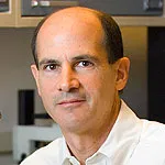
Photo by Steve Fisch of Dr. Thomas Rando.
Stanford Medicine News Center - May 30th, 2016 - by Krista Conger
There’s no place like home — particularly if you’re a muscle stem cell.
Snuggled comfortably along the length of our muscle fibers, these stem cells rest quietly, biding their time until the muscle needs to be repaired after injury. Although it’s possible to maintain muscle stem cells in a laboratory dish, they’re not really happy there. Within a short time they begin to divide and lose their ability to function as stem cells.
Now researchers at the Stanford University School of Medicine have come up with a way to create a home away from home for the stem cells in the form of artificial muscle fibers. They’ve also identified the particular “soup” of molecules and nutrients necessary to keep the cells in their most potent, regenerative state.
“Normally these stem cells like to cuddle right up against their native muscle fibers,” said Thomas Rando, MD, PhD, professor of neurology. “When we disrupt that interaction, the cells are activated and begin to divide and become less stemlike. But now we’ve designed an artificial substrate that, to the cells, looks, smells and feels like a real muscle fiber. When we also bathe these fibers in the appropriate factors, we find that the stem cells maintain high-potency and regenerative capacity.”
Why happiness matters
Keeping muscle stem cells happy in the lab is an important step toward potential therapies for conditions like muscular dystrophy and toward regenerating missing muscle after an injury. One day researchers would like to be able to remove a patient’s own muscle stem cells, correct any genetic deficiencies if necessary, and then transplant the cells back into the patient to regenerate healthy muscle tissues. This is not possible if the stem cells lose their ability to regenerate new muscle.
The researchers conducted most of their experiments in mice, using muscle stem cells from the animals. However, they were also able to show that human muscle stem cells remained more potent and could be more efficiently transplanted into laboratory mice when grown under similar conditions.
A paper describing the research was published online May 30 in Nature Biotechnology. Rando is the senior author. Former postdoctoral scholar Marco Quarta, PhD, is the lead author.
In order to prevent newly isolated muscle stem cells from activating when maintained in the laboratory, the researchers sought to identify genes whose expression increased when the cells begin to divide. Those genes, they reasoned, were likely to be involved in nudging the cells out of their potent, quiescent state and into a proliferative, less-stemlike state. Conversely, they also identified genes that were highly expressed in the quiescent cells.
Testing combinations of compounds
Once they had determined the gene-expression profiles unique to quiescence and activation, they tested various combinations of 50 compounds previously known or suspected to promote cell quiescence — adding them to the broth in which the cells were grown and watching whether the cells began to express genes involved with activation and to divide, or whether they continued to express the quiescence-associated genes. Eventually, the researchers came up with a panel of compounds that helped keep the cells potent over a period of about 48 hours.
Another key component of regenerative therapy is the ability of the quiescent stem cells to begin dividing after transplantation when they receive the appropriate triggers. Quarta and Rando found that growing the cells in the newly created broth for more than about three days compromised their ability to begin dividing when they were exposed to a combination of factors that normally promote growth. They speculated that the cells also needed the specialized environment of the muscle fiber to be optimally responsive to growth signals and to maintain their ability to reconstitute muscle tissue on demand.
A muscle fiber surrogate
Quarta and Rando joined forces with colleagues in Stanford’s Department of Materials Science and Engineering to figure out a way to assuage the homesick stem cells’ need for a muscle fiber to cling to. They needed the surrogate to be as close to the real thing as possible to prevent the cells from activating and losing their special stem cell properties. Ideally, it would have elasticity similar to real muscle fibers.
The researchers found that they could create elastic, artificial muscle fibers out of a naturally occurring, biocompatible molecule called collagen 1 by extruding it from a minipump to mimic the shape and geometry of a real muscle fiber.
“The process itself is quite simple,” said Rando. “The collagen extrusion device makes it easy and scalable. We can adjust the stiffness and size of the fibers, and then coat them with proteins we know are present in native muscle fibers.”
When the researchers applied muscle stem cells to the artificial collagen fibers, the cells quickly “homed” to places similar to those in which they would be found in real muscle. When maintained in the specially concocted quiescence broth, the cells appeared snug and happy along the collagen fibers. Furthermore, transplantation experiments indicated the muscle stem cells maintained their potency over several days and were able to quickly engraft and begin making new tissue in recipient animals.
The researchers also repeated the experiment using human muscle stem cells maintained under similar conditions and then transplanted into laboratory mice.
“This artificial niche, plus bathing the cells in the appropriate factors, allows us to maintain the cells in a highly potent, quiescent state,” said Rando. “Now we can genetically engineer stem cells, transplant them back into the animal and see that they are effective in engrafting and making new tissue.”
Other Stanford authors of the study are graduate student Jamie Brett; former graduate student Rebecca DiMarco, PhD; postdoctoral scholars Antoine De Morree, PhD, and James Su, PhD; former postdoctoral scholar Stephane Boutet, PhD; research associates Robert Chacon, Victor Garcia and Michael Gibbons; professor of cardiothoracic surgery Joseph Shrager, MD; and associate professor of materials science and engineering Sarah Heilshorn, PhD.
The research was supported by the Glenn Foundation for Medical Research, the National Institutes of Health (grants P01AG036695, R01AG23806, R01AR062185, R01AG047820 and EB001046), the California Institute for Regenerative Medicine and the Department of Veterans Affairs.
Stanford’s Department of Neurology and Neurological Sciences also supported the work.


