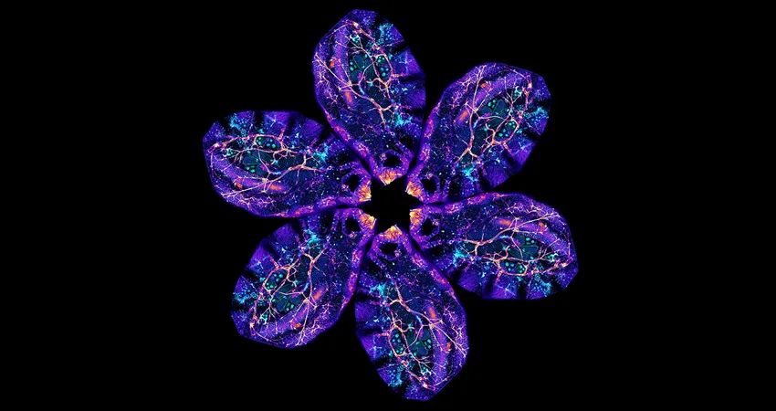
Image courtesy Lucy O'Brien.
January 27, 2020 - PLOS Biology
An innovative imaging platform by Lucy O’Brien, KC Huang, Andrés Aranda-Díaz, Leslie Koyama, and colleagues overcomes the challenge presented by the opacity of the adult Drosophila abdomen, enabling intravital imaging of live, intact flies at whole-organ and sub-cellular scales over multiple days.
Cell- and tissue-level processes often occur across days or weeks, but few imaging methods can capture such long timescales. The authors describe Bellymount, a simple, noninvasive method for longitudinal imaging of the Drosophila abdomen at subcellular resolution. Bellymounted animals remain live and intact, so the same individual can be imaged serially to yield vivid time series of multiday processes. This feature opens the door to longitudinal studies of Drosophila internal organs in their native context. Exploiting Bellymount’s capabilities, we track intestinal stem cell lineages and gut microbial colonization in single animals, revealing spatiotemporal dynamics undetectable by previously available methods.


