Stanford Report, August 15th, 2011 - by Andrew Myers
In 1991, Carl Lewis was both the fastest man on earth and a profound long jumper, perhaps the greatest track-and-field star of all time in the prime of his career. On June 14 of that year, however, Carl Lewis was human. Leroy Burrell blazed through the 100 meters, besting Lewis by a razor-thin margin of three-hundredths of a second. In the time it takes the shutter to capture a single frame of video, Lewis's 3-year-old world record was gone.
In a paper just published in the journal Neuron, a team at the Stanford School of Engineering, led by electrical engineers Krishna Shenoy and Maneesh Sahani, explored the neurological explanations for why Lewis may have lost that day. The team, which included graduate students Afsheen Afshar, Gopal Santhanam and Byron Yu and postdoctoral researcher Stephen Ryu, studied how the brain plans for and executes movements in reaction to a "go" signal.
New neurological measurement technologies and algorithms are allowing researchers a more complete look into how the brain functions. Engineers at Stanford are using these tools to better understand how the brain prepares to instruct the body to make a motion and why sometimes we react more quickly than others.
The advent of new measurement technologies that permit researchers to monitor up to hundreds of individual neurons simultaneously, combined with new analytical mathematics, are providing a revealing look inside the brain and a better understanding of the neurological processes behind the planning and execution of motion.
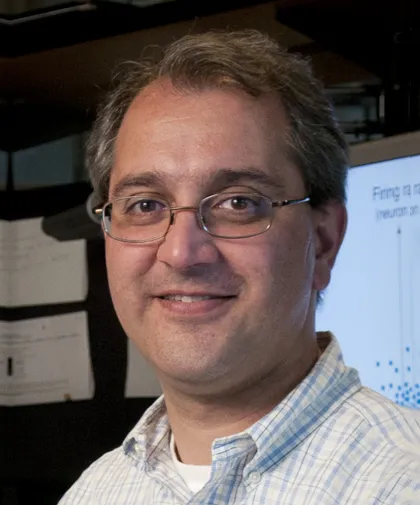
"This research holds great promise in many areas of neuroscience, in particular human prostheses that can be controlled by the brain," said Shenoy.
The ability of humans to time the onset of planned movements is imprecise, often frustratingly so. In Carl Lewis' case, that imprecision cost him the race and the record. In fact, experts later pointed out that Burrell was not really the faster man that day; he was merely the faster off the blocks, beating Lewis at the gun by about five one-hundredths of a second, a difference that provided the margin of victory.
"Lewis may well have lost because he wasn't able to optimize his own motor plan and thus his reaction time was slow," said Shenoy.
"Thanks to new tools, for the first time we are able to understand what the neurons are telling us," said Sahani. "We can hypothesize about how the activity of a group of neurons gives rise to movement."
Graduate students trained two rhesus monkeys to perform the task of touching a target on cue. The researchers then neurosurgically implanted on the surface of the monkeys' brains a 4-millimeter-square electronic chip arrayed with 100 tiny electrodes to monitor the neuron traffic.
The researchers concentrated on one particular area of the brain known as the dorsal pre-motor cortical area, which shows high levels of activity during the delay when arm movement planning takes place. Activity in this region varies depending upon the direction, distance and speed of a pending movement.
Where most historical data had been limited to single neurons, the new technology allows researchers to monitor in real-time the activity of hundreds of individual neurons down to the millisecond. They can now account for reaction times in single events, something previously impossible.
What Shenoy, Sahani and colleagues have found is a departure from the way many scientists had theorized the process worked. The existing hypothesis, known as "rise-to-threshold," held that in anticipation of a "go" cue, our brains begin to plan the motions necessary to satisfactorily complete the movement by simply increasing the activity of neurons.
Neurons begin to fire, but not enough to cause the movement to take place. Upon the "go" signal, the brain accelerates this neural firing until it crosses a "threshold" initiating the motion. According to the theory, the longer a preparatory period one has, the greater the neural activity will be and, thus, the faster the reaction time.
The Stanford team was able to document a process based less on the amount of activity and more on the trajectory of the neural activity through the brain. In graphs of neural activity prior to display of the target, the monkeys' neural activity appears somewhat scattered. The moment a target is displayed, however, the neural activity concentrates in an activity region that the researchers dubbed the "optimal sub-space."
"We can watch as the pattern of neural activity gets focused in a specific region at the moment the target appears," explained Shenoy, "and then when the 'go' cue is given, the activity moves again, ending with the successful touching of the target."
The key to reaction time, the researchers found, is the relationship between where the neural activity is and its speed along the ideal trajectory just prior to the go cue. If the neural activity is closer to the final destination, then the reaction time will be shorter; if farther away, then longer.
"We get our brains into a sort of ideal zone – an 'optimal space' – of neural activity," said Shenoy. "The planned movement is possible from anywhere within this space, but some points – those closer to the intended target along the ideal neural pathway – are more advantageous than others in terms of the reaction time."
From this new understanding, the researchers were able to shape a deeper understanding of the neural patterns and craft a model to predict reaction time.
"Our model allows us to predict with four times greater accuracy what the reaction time of any single arm motion is going to be, based on the neural activity observed prior to movement," said Sahani.
Returning to the practical applications, Shenoy and Sahani pointed to improving "neural prostheses" – artificial limbs and computer cursors that can be manipulated by the brain to help amputees and paralytics.
"A fundamental understanding of planning and movement is a central question in building electronic interfaces that convert neural activity into signals that can control computer cursors and prosthetic arms. These are also major areas of our research," said Shenoy.
This project was supported by the Collaborative Research in Computational Neuroscience (CRCNS) program – a joint initiative of the National Institutes of Health (NIH) and the National Science Foundation to support partnerships between experimental and computational neuroscientists. Afshar was supported by the NIH Medical Scientist Training Program, and Shenoy is funded by an NIH Director's Pioneer Award.
"This was a unique collaboration; Shenoy's team with its expertise in physiology and engineering and Sahani's expertise in computational modeling enabled them to take an innovative approach to understanding how the brain initiates movement. This research may ultimately have a significant impact on the development of neural prosthetics," said Yuan Liu, of NIH's National Institute of Neurological Disorders and Stroke, who was involved in the early development of the CRCNS program.
"For most of us, reaction times usually don't matter. Not many of us have to perform at the level of a Carl Lewis, after all," said Sahani. "But if you are an amputee hoping for a state-of-the-art prosthetic hand that you can control with your own brain, then understanding how the brain plans and executes motion is very important."