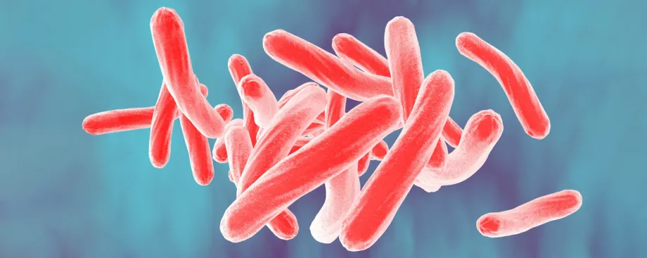
Illustration by Kateryna Kon, Shutterstock: An illustration of Mycobacterium tuberculosis bacteria.
Stanford Medicine News Center - March 8th, 2022 - by Krista Conger
An unexpected link between tuberculosis and cancer may lead to new drug treatments for the bacterial disease that kills more than 1.5 million people each year, according to a study led by researchers at Stanford Medicine.
The study found that lesions called granulomas in the lungs of people with active tuberculosis infections are packed with proteins known to tamp down the body’s immune response to cancer cells or infection. Some types of cancer drugs target these immunosuppressive proteins. Because these medications are widely used in cancer patients, the researchers expect that clinical trials can be launched quickly to test whether they can combat tuberculosis infection.
“This could be a paradigm shift in how people think about tuberculosis that has real, pragmatic implications about how to treat this disease,” said Mike Angelo, MD, PhD, assistant professor of pathology. “Our study suggests that unless you address the presence of these immunosuppressive proteins, you’re not going to get an effective recruitment of the immune system to fight the bacteria.”
Angelo is the senior author of the study, which was published Jan. 20 in Nature Immunology. Graduate student Erin McCaffrey is the lead author.
New tissue imaging technique
Angelo and McCaffrey used an imaging technique called multiplexed ion beam imaging by time of flight (MIBI-TOF), which was developed in the Angelo Lab in 2016. MIBI-TOF uses a mass spectrometer and specially tagged antibodies to track the locations of dozens of proteins within a tissue sample.
Angelo and his colleagues have recently used MIBI-TOF to identify the type and location of immune and cancer cells in tumors from people with triple-negative breast cancer— information that can be used to learn more about the biology and progression of these cancers.
When McCaffrey joined the lab, Angelo was on the lookout for other diseases that could be studied using the technique. “We try to focus on human diseases that manifest in solid tissue and seem to have remained intractable to current therapies,” Angelo said.
Tuberculosis fit the bill. The bacterial disease affects millions of people around the world, and it is difficult to cure even with extended antibiotic therapy. It’s also difficult to study in the laboratory because of a lack of suitable animal models. In addition, preserved samples of infected human lung tissue are relatively rare because the disease is not usually treated with surgery.
“Tuberculosis is a massive global health burden,” McCaffrey said. “Most of the time, the immune system is unsuccessful in eradicating the bacteria, but it’s not known why. We wondered whether the same molecular pathways that protect cancer cells from the immune system could also be affecting the immune response to the tuberculosis bacteria.”
The researchers used MIBI-TOF to map the location of immunosuppressive proteins — the same ones found in people with triple-negative breast cancer — in granulomas in lung and other tissue from 15 people with active TB. The results surprised them.
“We saw some of the brightest signals we’d ever seen, even in comparison with cancer tumors,” Angelo said. “This indicates the near-universal presence of key immunosuppressive proteins in the granulomas.”
In particular, the researchers saw high levels of two proteins — PD-L1 and IDO1 — that can suppress the immune response to cancer and are often found in tumor tissue. These proteins are targeted by approved cancer drugs.
When McCaffrey and Angelo studied blood samples collected from more than 1,500 people infected with TB, they found that the levels of PD-L1 correlated with clinical symptoms. Patients with latent, or asymptomatic, infection had lower levels of PD-L1 in their blood and were less likely to progress to active infection than those with higher PD-L1 levels. Conversely, patients with active infection who were considered cured after treatment experienced significant decreases in their blood PD-L1 levels compared with those who were not cured.
“We saw really consistent upregulation of these signals in the blood, which is emblematic of the failed immune reaction,” Angelo said. “They could also be used to predict disease progression from latent to active disease.”
T cells hang back
The researchers found that a type of immune cell in the granulomas called macrophages secrete high levels of PD-L1 and IDO1, as well as a protein called TGF-beta that also modulates the immune response. In addition, they found evidence suggesting that these macrophages are preventing another type of immune cell, the T cell, from being activated to fight the infection.
“This is different from what we see in cancer tumors, where the T cells are so active that they become exhausted and ineffective,” McCaffrey said. “Here, it looks like they’ve never been activated at all. This suggests that PD-L1 and IDO1 may be major players that prevent the immune response from ever getting off the ground.”
The researchers are homing in on the molecular pathways involved in this immune response to better understand how they might be reprogrammed to help the body eradicate the tuberculosis bacteria.
“The prevailing theory has been that tuberculosis is a disease enabled by T cell dysfunction,” Angelo said. “But now it seems like that isn’t the whole story. When taken at face value, this study suggests that macrophage dysfunction could be playing a superseding role that deters T cells from doing their jobs. If true, that is a totally different target for drug therapy, but the exciting part is that the tools we need to test this idea are already approved for use in humans.”
Other Stanford co-authors of the study are senior data scientist Michele Donato; graduate students Alea Delmastro and Noah Greenwald; postdoctoral scholars Sanjana Gupta, PhD, and Rashmi Kumar, PhD; former undergraduate student Alex Baranski; senior staff scientist Marc Bosse, PhD; life sciences technician Christine Camacho; pathology fellow Erna Forgo, MD; Sean Bendall, PhD, assistant professor of pathology; Matt van de Rijn, MD, professor of pathology; Niaz Banaei, MD, professor of pathology and of medicine; and Purvesh Khatri, PhD, associate professor of medicine.
Researchers from the Weizmann Institute of Science, Calico Life Sciences, the University of Wisconsin-Madison, the California Institute of Technology, the Africa Health Research Institute, the Texas Biomedical Research Institute, the Washington University School of Medicine, Emory University School of Medicine, the University of Texas Health Sciences Center and the University of Alabama at Birmingham are also co-authors of the study.
The research was supported by the National Science Foundation, the Damon Runyon Cancer Research Foundation, the National Institutes of Health (grants CA246880-01, R61/33AI138280, R01AI134810, 1U19AI109662, U19AI057229, 5R01AI125197, 5U54CA20997105, 5DP5OD01982205, 1R01CA24063801A1, 5R01AG06827902, 5UH3CA24663303, 5R01CA22952904, 1U24CA22430901, 5R01AG05791504 and 5R01AG05628705), CRDF Global, the South African Medical Research Council, the Department of Defense, the Wellcome Trust, the Parker Center for Cancer Immunotherapy, the Breast Cancer Research Foundation, and the Bill and Melinda Gates Foundation.


