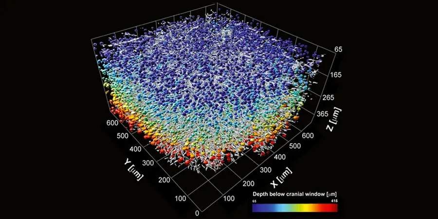
Visualization of a 3D CNS segmentation mask of brain neural networks in a living mouse at micron-level resolution, with a color-encoded depth of more than 10,000 CNS cell bodies, and their surrounding myelinated axons (grey).
July 30, 2022 - Nature Scientific Reports
The authors demonstrated 3D reconstruction of microarchitectural elements of a mouse cortex, and were able to visualize micro brain deformations and movements of neuron cells at a micron level. In the future, this technology could be used to visualize how the brain deforms over time as a response to injury or cancer growth, and highlight how the elasticity of the brain reacts to different treatments.
Optical coherence tomography (OCT) allows label-free, micron-scale 3D imaging of biological tissues’ fine structures with significant depth and large field-of-view. Here we introduce a novel OCT-based neuroimaging setting, accompanied by a feature segmentation algorithm, which enables rapid, accurate, and high-resolution in vivo imaging of 700 μm depth across the mouse cortex. Using a commercial OCT device, we demonstrate 3D reconstruction of microarchitectural elements through a cortical column. Our system is sensitive to structural and cellular changes at micron-scale resolution in vivo, such as those from injury or disease. Therefore, it can serve as a tool to visualize and quantify spatiotemporal brain elasticity patterns. This highly transformative and versatile platform allows accurate investigation of brain cellular architectural changes by quantifying features such as brain cell bodies’ density, volume, and average distance to the nearest cell. Hence, it may assist in longitudinal studies of microstructural tissue alteration in aging, injury, or disease in a living rodent brain.