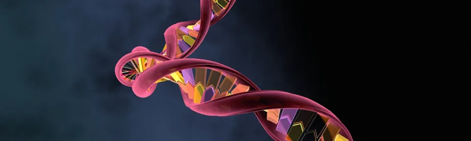
Graphic image by Motionstream, Shutterstock. Watch video!
Stanford Medicine Scope - May 12th, 2016 - by Krista Conger
Deciphering the shape of living molecules inside a cell presents a conundrum. Often the very process of preparing the molecule for study destroys the structures scientists most wish to see. This engaging video outlines a clever new technique devised in the laboratory of genome scientist Howard Chang, MD, PhD, to determine the three-dimensional structure of RNA molecules. Some of these carry the instructions to make critical proteins in the cell; others (known as long, non-coding RNAs or lncRNAs) serve as regulatory molecules to control other cellular functions. Chang and his colleagues published their work today in Cell.
As Chang explained to me:
RNA molecules can fold up into complex structures, much like how paper can be folded to make complex origami shapes — a bird, a boat, or a flower. The RNA shapes determine their function, but it has been difficult to see the three-dimensional shapes of RNA inside cells. In the past, scientists relied mostly on one-dimensional methods; although they could tell which part of the RNA was single-stranded or double-stranded (paired), but we did not know for sure which double-stranded parts in a molecule paired together.
Postdoctoral scholars Zhipeng Lu, PhD, and Qiangfeng Cliff Zhang, PhD, figured out a way to temporarily bind together the two strands of paired RNA within a living cell using a chemical called psoralen. They then isolated the short, double-stranded structure, attached a short hinge section, and reversed the psoralen binding. The result is a single-stranded RNA molecule, each half of which represents a partner in a bonded pair. The technique, which the researchers termed PARIS (for psoralen analysis of RNA interactions and structures) yielded some fascinating insights into what they termed the “structurome” and the “interactome” that make up the hidden lives of RNA molecules.
As Chang described:
Many previous strategies for analyzing double-stranded RNA structures have assumed that this base pairing would most likely occur over short distances between closely neighboring nucleotides. But we discovered that many of these pairs occur over thousands of nucleotides within single molecule. In fact, it’s even possible for the beginning and end of a single molecule to pair, making a circle. This could be very advantageous for RNA molecules that need to be highly translated, because the protein-making ribosomes can just go around and around without needing to release the molecule and go back to the beginning.
The researchers also found that many RNA structures are evolutionarily conserved among species and that single RNA molecules often assume a variety of conformations within the cell, possibly acting as reversible molecular switches. Using the technique helped solve a long-standing mystery as to the shape of an important lncRNA called XIST that’s been a particular research focus in Chang’s lab.
“We’ve just begun to scratch the surface of what we can learn with this technique,” Chang told me. “It’s broadly applicable to RNA structures in any organism: plants, bacteria, pathogens and more. Now we can learn the higher-order structures of RNA within living cells, and it’s very exciting.”
