
Photo by Norbert von der Groeben - Adam de la Zerda developed a technique known as photoacoustic molecular imaging, which allows researchers to see cancerous tumors hiding under tissues.
Inside Stanford Medicine - April 8, 2013 - by Elizabeth Devitt
When Adam de la Zerda commuted home from his postdoctoral chemistry work at UC-Berkeley, he often stopped at a French restaurant in San Mateo. But he didn't join the other diners. Instead, he headed for the kitchen where he swapped his lab books for cookbooks and stood in as a sous chef.
"There's a lot of chemistry in cooking," said de la Zerda. His favorite culinary creation was quail eggs and caviar, with the eggs cooked "sous vide" style, a technique that vacuum-seals food in bags and then slow cooks the meal in water. In consultation with the chef, de la Zerda perfected the dish by calculating tissue deterioration rates to find the ideal incubation time.
Managing a spot as a sometime-sous chef isn't anyone's typical pastime, but it's characteristic of de la Zerda. The 28-year-old faculty member, recently appointed an assistant professor in the Department of Structural Biology, has a passion for interdisciplinary pursuits.
Originally from Israel, de la Zerda was "dead set" on getting his degree in theoretical quantum physics when he applied for graduate school in electrical engineering at Stanford. That was the logical next step after an undergraduate focus on computer engineering and physics. But, when he saw an engineering friend choose an adviser from the Music Department, de la Zerda suddenly appreciated he was at a university that didn't dwell on departmental divisions. "When I realized that, I thought: 'What else can I do?'"
De la Zerda's new technique uses nanoparticle agents to convert light waves into ultrasound waves, and then generates more informative images than methods such as computed tomography or magnetic resonance imaging.
Spurred by the loss of a good friend to cancer, de la Zerda wanted to improve treatment strategies. He knew one big problem was the lag time in current imaging technology — often two to three months — between starting chemotherapy and finding out whether a tumor was shrinking. Before tumors visibly change, there's molecular activity going on; proteins are being shut down and other abnormal activities occur that could show whether the tumor was responding to treatment. "If we could only see those molecular activities, doctors could quickly tell which patients are not responding and prescribe a different treatment," he said.
To explore new solutions, de la Zerda joined the lab of Sam Gambhir, MD, PhD, professor and chair of radiology and director of the Molecular Imaging Program at Stanford. There, he developed and patented a technology named photoacoustic molecular imaging under the guidance of Gambhir and in collaboration with Pierre Khuri-Yakub, PhD, professor of electrical engineering. This new technique uses nanoparticle agents to convert light waves into ultrasound waves, and then generates more informative images than methods such as computed tomography or magnetic resonance imaging. With nanoparticles targeted at biomarkers in a tumor, those images can show specific molecular activity in the tissue.
The unique advantage of photoacoustic molecular imaging is that it allows scientists to see tumors hiding under other tissues and structures. It can also outline tumor boundaries during surgery, which helps surgeons see what to cut out and what to leave in — avoiding mistakes either way. "It's like having Superman vision," said de la Zerda.
"There may be a million different things we can do with this," he added. "We can study basic tumor biology. We can monitor the treatment of cancer patients. We can even apply this technique to diseases other than cancer."
It turns out this technique is particularly useful in diseases such as age-related macular degeneration, an eye disease in which the central field of vision is lost. It's hard to detect early changes to the health of light-sensing cells at the back of the eye. But, with co-investigator Mark Blumenkranz, MD, professor and chair of ophthalmology, de la Zerda refined the photoacoustic imaging. He designed a way to look at growth of new blood vessels under the retina, a hallmark of the "wet" form of the disease, without touching the inside of the eye. This preliminary study led to a Bio-X program seed grant as part of its Interdisciplinary Initiative Program.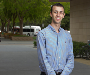
The potential of de la Zerda's work attracted the attention of faculty in the Department of Structural Biology. "Adam has a combination of mind and personality and obvious energy that stand out," said Roger Kornberg, PhD, a professor of structural biology and a Nobel laureate in chemistry, who was one of the department members who unanimously decided to appoint de la Zerda to its faculty.
Imaging has captivated de la Zerda since third grade, when an aunt gave him a small microscope. "I remember it like it was yesterday," he said of the early foray into science. "I collected dead bees, and under the microscope lens I could see they were made of amazing and beautiful structures. My dad would explain how structure correlated to their function; how all the angles of their eyes helped them see. I would see things that I couldn't see just by looking with my own eyes, and that fascinated me."
Although he's made his first marks with imaging, de la Zerda said he's an engineer at heart: "For me, it's important that someone can use the tools I'm building. If no one ends up using them, I might as well have never built them." The satisfaction of turning his ideas into an entity that could help someone also lured him into the arena of Silicon Valley startups. With a modest seed investment, he co-founded OcuBell, a company dedicated to improving ophthalmic imaging options.
But it isn't just building tools that interests de la Zerda; he also wants to help others bridge the gap between engineering and medicine. "I want electrical engineers and materials scientists to know they can tackle some of the most important problems in biology and medicine," he said. So, he created a graduate-level class — "Bio-chips, Imaging and Nanomedicine" — in collaboration with Shan Wang, PhD, professor of electrical engineering and of materials science and engineering, and Demir Akin, DVM, PhD, deputy director of the Center for Cancer Nanotechnology Excellence, to provide engineering students with foundations in human biology and medical diagnostics.
The first class was a hit, attracting more than 100 students. Now in its third year, the course has almost 90 students enrolled.
"It wasn't my blue eyes that brought students to class," de la Zerda said. "They came because they were honestly curious for how their engineering background could be applied to biology and medicine.
"Stanford is a very unique place as people do not classify you only for your accomplishments, but also for where your passion is," he said. "It's almost a mission" to tell other students to reach beyond what they think they know, he added. "I want them to know the sky's the limit here."
Although he's only been on the Stanford faculty since August, de la Zerda's wide-ranging efforts have garnered numerous awards. Most recently, he was listed in Forbes magazine "30 under 30" in science and health care and received the Dale F. Frey Award from the Damon Runyon Cancer Research Foundation, as well as the Director's Early Independence Award from the National Institutes of Health.
While he appreciates the accolades and attention such honors bring to his work, de la Zerda prefers to talk about the encouraging environment at Stanford.
"Here, all things are possible," he said.
