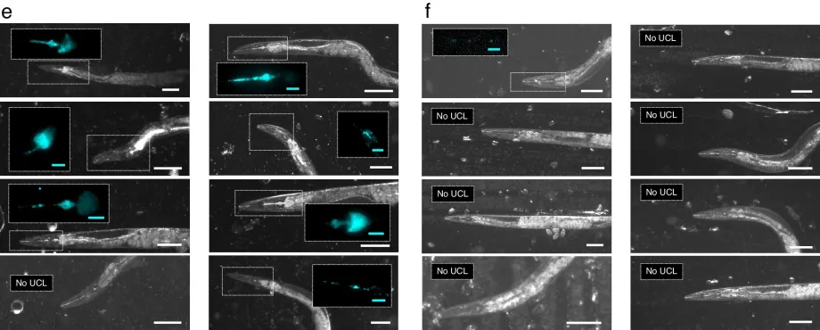
A series of bright field images of levamisole-treated worms after one-hour incubation with e, microgauges or f, singly dispersed PMAO-wrapped UCNPs. Scale bars are each 100 µm. False-colored UCL images (cyan) of the boxed pharyngeal regions are displayed in the inset of each picture, when applicable. Inset scale bars are each 20 µm each. The anterior of each animal is to the left.
Stanford Bio-X affiliated faculty members Miriam Goodman, Jennifer Dionne, and Wendy Gu, with co-authors Stanford Bio-X Travel Award recipients Cindy Shi and Ariel Stiber and first author Jason R. Casar have developed a nanoparticle technique that can be used to measure force dynamics inside a living creature, such as Caenorhabditis elegans worms biting their food.
In their paper published in the journal Nature, the group describes how they used infrared radiation to excite luminescent nanocrystals in a way that allowed the energy levels of cells inside a C. elegans worm to be measured.
being able to measure forces inside living creatures could go a long way toward a better understanding of internal molecular processes. Such efforts have thus far been stymied by the need for remote sensing over variable ranges and scales. In this new study, the research team has found a way around such problems, and they have measured the force of the muscles inside the digestive tract of C. elegans.
READ MORE in Nature READ MORE at Phys.Org READ MORE in Nature News and Views