Stanford Report - April 15th, 2009 - by Krista Conger
A drop of blood or a chunk of tissue smaller than the period at the end of this sentence may one day be all that is necessary to diagnose cancers and assess their response to treatment, say medical school researchers.
In a study published April 12 in the online version of Nature Medicine, the scientists used a specialized machine capable of analyzing whether individual cancer-associated proteins were present in the tiny samples and even whether modifications of the proteins varied in response to cancer treatments. Although the study focuses on blood cancers, the hope is that the technique might also provide a faster, less invasive way to track solid tumors.
Stanford scientists used a specialized machine capable of analyzing whether individual cancer-associated proteins were present in the tiny samples and even whether modifications of the proteins varied in response to cancer treatments - the technique might also provide a faster, less invasive way to track solid tumors.
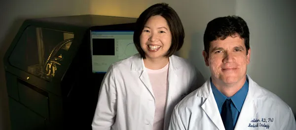
"Currently we don't know what's going on in a patient's tumor cells when a treatment is given," said Alice Fan, MD, a clinical instructor in the division of oncology. "The standard way we measure if a treatment is working is to wait several weeks to see if the tumor mass shrinks. It would be a leap forward if we could detect what is happening at a cellular level." Fan, the lead author of the study, performed the research in the lab of senior author Dean Felsher, MD, PhD, associate professor of medicine and of pathology and the leader of the Stanford Molecular Therapeutics Program.
"This technology allows us to analyze cancer-associated proteins on a very small scale," said Felsher, a member of Stanford's Cancer Center, who studies how cancer genes called oncogenes initiate and influence tumor progression. "Not only can we detect picogram levels—one-trillionth of a gram—of protein, but we can also see very subtle changes in the ways the protein is modified."
Variations in the way a protein is modified, or phosphorylated, can affect how it functions in tumor progression. Cancer cells often evade common therapies by rejiggering their levels of protein expression and degrees of phosphorylation. Analyzing repeated small samples from a tumor undergoing treatment may let doctors head off rogue cells at the pass before they proliferate into a more resistant tumor as well as identify patients likely to fail standard approaches to treatment.
Fan and Felsher collaborated with researchers from Palo Alto-based Cell Biosciences, which makes the machine used in this study, to separate cancer-associated proteins in narrow capillary tubes based on their charge, which varies according to modifications on the proteins' surfaces. Two versions of the same protein—one phosphorylated and one not—can be easily distinguished because they travel different distances in the tube. The researchers then used antibodies to identify the relative amounts and positions of the various proteins.
Not only was the technique able to identify oncogene activation in cultured tumor cells, but it also worked in lymphoma samples drawn from mice with small, hollow needles. Furthermore, it was able to detect varying levels of expression of two common oncogenes in 44 of 49 lymphoma samples from human patients as compared with normal controls, and even distinguish some types of lymphomas from others.
Finally, it could detect subtle differences in phosphorylation in several other cancer-associated proteins. "Some of these proteins can exist as five or six phosphorylated variants," said Felsher. "With this technology we can see changes that occur in as little as 10 percent of the total protein pool. Now we have a tool that will help us look at what's happening in cells over time."
"Surgical biopsies usually require general anesthesia and large amounts of tissue," agreed Fan. "If we can figure out how to go in with a needle and remove just a few cells for analysis, we could repeatedly assess how the tumor is responding to treatment."
For example, the researchers were able to confirm through serial biopsies of a human lymphoma patient that, as suggested by previous research in the Felsher lab, the lipid-lowering drug atorvastatin reduces phosphorylation of yet another cancer-associated protein. "This is the first time we've been able to see that this compound affects the biology of cancer cells in patients," said Felsher.
Fan is expanding her investigations to include head and neck tumors, which tend to be accessible for cell sampling. Fan and Felsher caution that more research is needed before the technology would be widely available clinically.
The study also involved visiting medical student Mathias Orban; Jason Gotlib, MD, assistant professor of medicine; Yasodha Natkunam, MD, associate professor of pathology; research associate Liwen Xu, PhD; study coordinator Amy Shirer, MD; and research assistant Ada Yee. It was supported by the National Cancer Institute, the Burroughs Welcome Fund, the Damon Runyon Foundation and the Leukemia and Lymphoma Society.
 and of Pathology")