Inside Stanford Medicine - November 8th, 2010 - by John Sanford
The Nuclear Medicine and Molecular Imaging Clinic that opened last month at Stanford Hospital & Clinics will help advance a new generation of diagnostic techniques for earlier detection and improved management of cancer, heart disease and neurological disorders.
The $25 million clinic is outfitted with the latest equipment, including two new PET/CT scanners and a new cardiac scanner that produces a three-dimensional image of the blood supply to the heart in only 4-6 minutes, compared with the 20-24 minutes it took the old machine. The clinic also will house developing technologies that promise to revolutionize medical imaging.
“We’ll be able to see more patients more quickly,” says Gambhir. “We can also ramp up clinical studies to get a better understanding of, for example, neurological disorders like Parkinson’s disease, Alzheimer’s and epilepsy.”
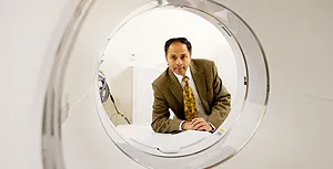
“It’s designed to serve patients as well as support clinical trials,” said Sanjiv “Sam” Gambhir, MD, PhD, who heads the hospital’s Division of Nuclear Medicine and the Molecular Imaging Program at the School of Medicine. “We’ll be able to see more patients more quickly. We can also ramp up clinical studies to get a better understanding of, for example, neurological disorders like Parkinson’s disease, Alzheimer’s and epilepsy.”
The clinic’s new PET/CT scanners are state-of-the-art, said Jayesh Patel, technical manager of nuclear medicine, and “can be used to detect miniscule evidence of disease.”
Computerized tomography uses X-rays to create an image of the structure of organs and tissue. Positron- emission tomography is a type of nuclear medicine that uses a scanner and a radioactive isotope injected into patients to measure how well particular bodily functions, such as blood flow, molecular receptors and sugar metabolism, are working.
The clinic occupies 16,000 square feet on the second floor of Stanford Hospital, just above the Market Square Cafeteria. With several scanning rooms, laboratories, doctors’ offices and a central control room for technologists, it brings together important imaging and lab functions that were previously dispersed across the medical center. This consolidation means patients can have molecular-imaging work done at one time and in one place, Gambhir said, and will likely speed up how quickly they learn about their diagnoses.
The clinic’s previous location, a 6,000-square-foot space on the hospital’s lower level, will house other imaging equipment, including a new MRI machine.
Gambhir is one of the leading researchers in molecular imaging, which uses nuclear medicine to understand how organs and tissue operate at the cellular and subcellular levels. The clinic will be a valuable resource for a number of his team’s research efforts in this area. One of the most promising, Gambhir said, is in vitro diagnostics, in which samples of blood and tissue are scanned or otherwise tested at the molecular level and then compared with in vivo imaging — that is, real-time images of patients.
The big advantage of test-tube samples, Gambhir said, is that they can be obtained more frequently than a patient can be imaged. “You can’t subject someone to PET or CT every couple of days, but a person can provide a blood sample every few days that we can analyze with new diagnostic tests,” Gambhir said. This way, the progress of a disease can be closely followed, and physicians can better determine whether a particular treatment is working, he said.
“We think the future of early detection monitoring and patient disease management in general is a combination of in vitro and in vivo diagnostics,” he added. “It’s like a merger of pathology and radiology.”
Optical scanners that can detect breast tumors using only light — not X-rays, which may have some side effects due to radiation — are also going to be used in clinical trials, Gambhir said. Optical scanners use lasers to look for tumors by measuring how much light emerges from the other side of scanned tissue. The lasers also can be used to illuminate imaging agents that have been injected into a patient and designed to lock on to tumors. Gambhir said he believes optical scanners will likely replace X-rays in the future as the preferred technique for mammograms.
In addition, Gambhir and his team hope to use photoacoustic tomography to look for cancer in patients. This technique involves targeting tumors with lasers that lead to light absorption in the body and the production of sound waves. “This method can give you excellent depth penetration,” Gambhir said. “The light can be very precisely targeted, but sound penetrates through tissue much better and gives you a better spatial resolution.”
The scientists at the Canary Center at Stanford for Early Cancer Detection, which Gambhir also directs, will take advantage of the new clinic as they work on new methods to identify cancers and then translate their research into clinical trials and, ultimately, into general practice. “The Canary Center and this new clinic will go hand-in-hand,” Gambhir said.
