Graphic by nobeastsofierce, Shutterstock.
Stanford Medicine Scope - June 26th, 2017 - by Nathan Collins
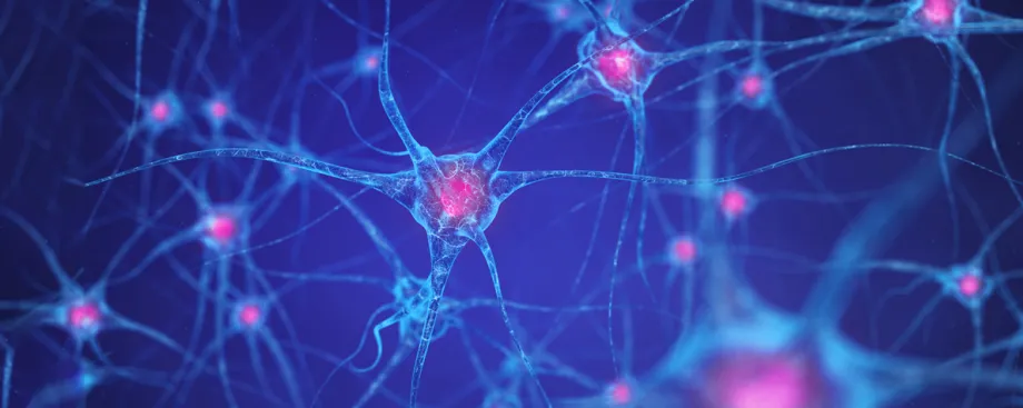
Imagine being able to take a crystal-clear snapshot of an entire brain, recording what every single neuron was doing at a particular moment as an animal experienced fear or pleasure or any other emotion. Today, that’s just a dream — neuroscientists have to choose between seeing the entire brain in low resolution or seeing a small piece of it in high resolution — but a new technique known as FLARE could bring that dream one step closer to reality.
The research emerged, says Alice Ting, PhD, out of neuroscientists’ frustration with their inability to capture a fine-grained picture of what the whole brain was doing in experiments, although some well-timed “pestering” also played a role. Ting says her friend and collaborator Kay Tye, PhD, a neuroscientist at the Massachusetts Institute of Technology, kept asking her to develop a method that could achieve both a fine resolution and a wide scope.
Eventually Ting, a professor of genetics and biology and a member of Stanford Bio-X, acquiesced, partly because she had hired a postdoc, Wenjing Wang, PhD, who she thought was up to the challenge.
“Wenjing is very fearless, [and] this project could only even be attempted by someone who is utterly fearless, because the idea was a little bit crazy,” Ting said.
Crazy, she explains, because they wanted to solve several problems at once. First, they wanted to create a snapshot of the entire brain, or at least a very large number of neurons, all at once and in great detail. Second, they wanted that snapshot to single out a short window of time for analysis — for example, a window during which mice react to sounds or experience fear. Finally, the team wanted a method that would allow them to not just image the brain, but also go back in and dial neural activity up or down.
FLARE, which stands for Fast Light- and Activity-Regulated Expression, was the solution. The research describing the work appears today in Nature Biotechnology. The method works by tagging neurons that are firing with a molecule that glows. Crucially, the tagging only happens when neurons are exposed to light, allowing researchers to focus on brain activity during a window of time they select simply by turning a light on and off again.
Three features of FLARE distinguish it from existing technologies, Ting says. First, because the tagging process itself is permanent and works in all active neurons, the entire brain becomes a permanent record of neural activity, one that researchers can essentially scan into a computer and revisit whenever they like. (The caveat is that to construct a picture of all of those neurons at once, one has to remove the brain, unfold it, and go over each little piece with a microscope — so don’t expect this to work in people anytime soon.)
Second, FLARE’s time resolution is a vast improvement over other methods that create similar permanent records. Previously, a snapshot took about 12 hours or more to make, making it impossible to single out brain function during just one activity or behavior. FLARE images, in contrast, take just a few minutes to make. Future versions could bring that number down to seconds, Ting said.
Finally, the tag itself is modular, meaning that researchers can attach nearly whatever they want to it — in particular, they could activate or deactivate any gene that want, allowing neuroscientists to trigger fluorescence, dial neural activity up or down to treat depression, or, Ting said, pretty much anything else.
“It’s quite different from nearest neighbor technologies, and opens up new possibilities for neuroscientists,” Ting said.
Wang describes her project in somewhat simpler terms: “It’s really awesome.”