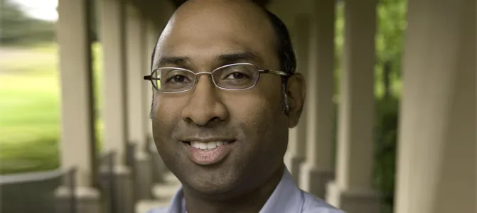
Dr. Ravindra Majeti.
Stanford Medicine Scope - May 24th, 2016 - by Christopher Vaughan
In the history of science and medicine, the breakthrough discoveries get a lot of deserved attention, but often overlooked are the invention of the tools that made those discoveries possible. Galileo discovered moons around other planets, but it was the invention of the telescope that made his observation possible. Leeuwenhoek discovered microbes only after he created a simple but powerful microscope.
Researchers in the laboratory of Stanford’s Ravi Majeti, MD, PhD, have just created one such tool — a method of implanting cells to create a human bone-like structure in mice. This allows investigators to study a whole host of blood cancers and other diseases that they had trouble studying before. Majeti and colleagues published the work this week in the journal Nature Medicine.
“Transplanting human leukemia cells into mice has been an important way of studying the disease,” postdoctoral fellow Andreas Reinisch, MD, PhD, first author of the paper, recently explained. “This method has given us important insights into how cancer develops and has been the way that all new anti-leukemia drugs have been tested and developed.” Mice engrafted with human leukemia cells are even sometimes used to test possible drug treatments for patients currently undergoing treatment for the blood cancer, he added.
The problem, said Reinisch, is that even in the best cases, researchers can get human leukemia to take up residence and multiply in mice only half the time. For many types of blood cancers, engraftment doesn’t happen at all. It’s like Galileo’s telescope could only be pointed at a small spot of sky visible through a hole in his roof.
Majeti, Reinisch, and their co-workers have metaphorically blown the roof off by transplanting special bone marrow-derived cells called stromal cells in mice. These human stromal cells multiply and divide to create a miniature, bone-like structure, complete with a bony outer shell and an internal, marrow-like compartment. This structure allows implanted human blood cells to live and grow in their natural environment — human bone.
“I’m really excited about this technique because we now have a way to look closely at all kinds of leukemia, as well as many other profilerative disorders of blood cells,” Majeti said. “It also opens up the study of other, non-blood diseases such as metastatic breast cancer, which often spreads to bone in humans but simply won’t spread in existing mouse models.”