Graphic by AuntSpray, Shutterstock.
Stanford Medicine Scope - June 29th, 2017 - by Erin Digitale
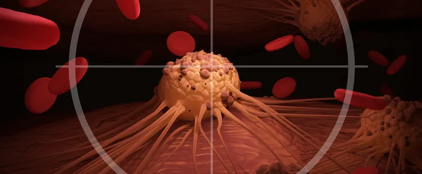
Molecular scissors sticking out from the surface of a hard-to-treat type of brain tumor could be harnessed to deliver drugs to exactly the right spot in the brain, a new Stanford study suggests.
In the study, published this week in Molecular Cancer Therapeutics, a team led by pediatric radiologist Heike Daldrup-Link, MD, tried a new strategy to tackle a brain tumor called glioblastoma. Glioblastoma is a particularly awful tumor — the mean survival time after diagnosis is now 12 months in both children and adults. Daldrup-Link’s research was conducted in mice implanted with human glioblastoma tumors; the findings still need to be confirmed in people.
The tumor is difficult to treat because a small group of aggressively malignant cells, called glioblastoma-initiating cells, are very good at hiding from existing forms of chemotherapy. When glioblastoma is surgically removed and treated with radiation and chemotherapy (the standard approach now), it recurs in 90 percent of patients. Researchers think GICs start the regrowth.
But these nasty cells have a potential weakness: They rely on tumor blood vessels to survive. If these blood vessels can be destroyed, researchers think the GICs will die, too.
Tumor blood vessels are leakier and less organized-looking than normal blood vessels, and some prior attempts to get chemo drugs into tumors have relied on the resulting “enhanced permeability and retention effect” — basically, the idea that drugs will leak into tumors more easily than into healthy tissue. But this leaky-pipe effect isn’t uniform enough to reliably wipe out GICs.
Hence the Stanford team’s idea. They decided to try harnessing a different property of tumor tissue. Many copies of an enzyme called matrix metalloproteinase-14 stick out from glioblastoma cells. These enzyme molecules function as high-specificity scissors, able to snip a certain sequence of protein in two.
To take advantage of the enzyme, the scientists built a sort of molecular Tinkertoy with three components: a cancer-drug precursor at one end, an iron nanoparticle at the other and a connector in the middle consisting of the protein targeted by the molecular scissors.
Early tests in a different tumor model suggested this system worked as the researchers hoped. The cancer enzyme snipped the middle of the “Tinkertoy”, which the researchers call CLIO-ICT, in two, releasing and activating the cancer drug exactly where it was needed.
In the new study, the team showed that their strategy helped fight glioblastoma. The drug caused collapse of tumor blood vessels, appeared to starve GICs (the very nasty cells) and slowed tumor growth. There was another advantage, too: After the molecular scissors did their job and released the cancer drug, the iron nanoparticles from the other end of CLIO-ICT hung around the tumor and showed up on MRI scans. The iron helped researchers monitor the shrinkage of the tumor.
In the new study in mice, the scientists also confirmed that CLIO-ICT doesn’t hurt healthy organs such as the heart and lungs. In these healthy organs, absent the cancer enzyme, the cancer drug isn’t released or activated, preventing possible toxicity. And CLIO-ICT worked even better when given in combination with a second cancer drug, temozolomide, that is already used to treat glioblastoma.
The scientists hope their new approach will ultimately be used to improve survival in people with the brain tumor.


