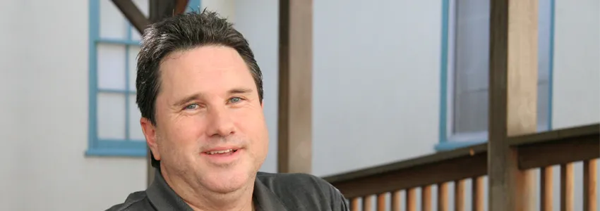
Ron Blackwell took part in a procedure at Stanford Hospital & Clinics in which electrodes were temporarily implanted in his brain. When two nerve clusters received mild stimulation, Blackwell's perception of a researcher's face instantly warped and changed. Photo by Jonathan Rabinovitz.
Inside Stanford Medicine - October 23, 2012 - by Bruce Goldman
In a painless clinical procedure performed on a patient with electrodes temporarily implanted in his brain, Stanford University doctors pinpointed two nerve clusters that are critical for face perception. The findings could have practical value in treating people with prosopagnosia — the inability to distinguish one face from another — as well in gaining an understanding of why some of us are so much better than others at recognizing and remembering faces.
In a study published Oct. 24 in the Journal of Neuroscience, the scientists showed that mild electrical stimulation of two nerve clusters spaced a half-inch apart in a brain structure called the fusiform gyrus caused the subject’s perception of faces to instantly become distorted while leaving his perception of other body parts and inanimate objects unchanged.
The surprised reaction of the subject, Ron Blackwell of Santa Clara, Calif., is captured in a video made during the procedure. “You just turned into somebody else. Your face metamorphosed,” he tells the researcher in the video.
Blackwell, who is now 47, was undergoing medical treatment under the direction of Josef Parvizi, MD, PhD, associate professor of neurology and neurological sciences at the School of Medicine, whose lab collaborates with that of Kalanit Grill-Spector, PhD, associate professor of psychology at the School of Humanities and Sciences.
The face Blackwell was looking at was Parvizi’s. “Ron didn’t see my face vaporize or go blank. Instead, it just seemed to warp before his eyes,” Parvizi said.
In 2010, Grill-Spector and then-graduate student Kevin Weiner (now a postdoctoral researcher in Grill-Spector’s lab and a co-author of the new study) discovered that the fusiform gyrus contains two nerve clusters (designated as pFus and mFus) that respond more strongly to faces than to hands, legs, cars, guitars, flowers or buildings.
Grill-Spector’s lab has been studying the fusiform gyrus’ role in face recognition as well as in prosopagnosia or “face blindness,” a condition made famous by neurologist and author Oliver Sacks, MD, who himself suffers from it. People with prosopagnosia simply cannot distinguish one face from another, although all other aspects of their vision and visual-information processing are normal. Some people, like Sacks, are born with the condition while others acquire it as a result of an injury to the fusiform gyrus, Grill-Spector said.
“We can learn a lot about the function of different brain regions by studying these disorders and relating them to the anatomical sites where brain damage has occurred,” she said. “But the injuries vary a great deal from one affected person to the next, and they are typically not confined to the fusiform gyrus. This limits our ability to localize a particular deficit to a particular brain site.”
Blackwell’s arrival at Stanford Hospital & Clinics, along with a generous slice of serendipity, allowed the Stanford doctors to conduct the first-ever study of the fusiform gyrus combining two imaging techniques (fMRI and electrocorticography, or intracranial recording) and electrical brain stimulation. The result was the first-ever proof that appropriate activity in the two nerve clusters, pFus and mFus, was critical to face recognition.
Blackwell had time on his hands. He was spending a week in a bed at Stanford Hospital with a packet of electrodes snugly abutting a part of his brain that doctors believed might be the initiating site, or focus, of his epileptic seizures. These seizures, which he’d been experiencing since age 11, had been well-controlled by drugs. But by late 2010, his medication was failing.
Blackwell was married and the father of two young girls. “It wasn’t just me anymore. I didn’t want to be vulnerable to disorienting episodes that could occur at any time of the day or night,” he said. Referred by his primary physician to Parvizi, he learned of a medical procedure that might provide relief.
Epileptic seizures are, at root, electrical storms triggered when a short circuit at one small spot within the brain, called the focus, causes waves of electrical activity to spread throughout the organ. (The exact location varies from patient to patient.) Most of the time seizures can be controlled with medication. When they can’t, one proven treatment involves a surgical procedure in which a portion of the patient’s skull is temporarily removed, allowing access to the surface of the brain near the spot thought to be responsible for initiating the seizures. A packet containing numerous electrode leads is placed near the brain’s surface, with each electrode acting like a separate stethoscope monitoring the collective electrical activity of perhaps a half-million nerve cells. (That’s a drop in the bucket, considering that a healthy human brain contains 200 billion of them.) The patient remains off medication for several days, eventually culminating in the onset of seizure activity and allowing the neurological team to identify the focus. Surgeons may then be able to excise just enough brain tissue to halt the cycle of self-propagating electrical activity — in effect, pulling out a fuse in order to break the short circuit — without affecting any important brain functions.
In September 2011, Blackwell was set to undergo the weeklong monitoring procedure. Based on a thorough neurological workup, electrodes were placed on the surface of a brain region that included the fusiform gyrus, a structure roughly the shape of a hand-rolled cigarette, on the underside of the temporal lobe.
Electrical brain stimulation requires the flow of electricity from one electrode across a small patch of brain tissue to another electrode spaced 1 cm away. It so happens that pFus and mFus, which account for perhaps one-quarter of the fusiform gyrus’ bulk, are also 1 cm apart. By fate, two of the inserted electrodes had been positioned almost precisely above the anatomical centers of pFus and mFus in Blackwell’s fusiform gyrus, forming a pair that enabled Parvizi to apply the stimulation directly to the two sites simultaneously at the touch of a button.
Doing so instantly altered Blackwell’s perception of Parvizi’s face. “You almost look like somebody I’ve seen before, but somebody different,” he reported. “... You were someone else. Your whole face just sort of metamorphosed ... it’s almost like the shape of your facial features drooped.”
When the stimulation was halted, the distorted image of Parvizi’s face immediately reverted to the normal one, Blackwell reported. Neither sham stimulation, in which a button was pushed but no actual electrical impulse was delivered to pFus and mFus, nor stimulation of nearby sites via other electrode pairs had any effect on Blackwell’s face perception. Stimulating pFus and mFus caused no distortion of other objects in the room, he reported.
Throughout this procedure’s duration, Parvizi didn’t know what to expect. “I was as surprised as the patient was when he suddenly saw my features seem to melt,” Parvizi said.
In a follow-on imaging study of Blackwell’s brain using a high-resolution fMRI technique that could discriminate among locations as little as 1.8 mm apart, Grill-Spector and her students confirmed Parvizi’s findings. Blackwell’s pFus and mFus clusters responded with heightened activity to images of faces, but not to those of hands, feet, flowers, cars, guitars, cars or houses.
The Stanford team of neurologists succeeded in locating the focus of Blackwell’s seizures, but decided that removing it would be risky because it was too close to other key parts of the brain. But oddly, since the removal of the electrodes, his medication seems to be working again and his seizures have inexplicably abated to a significant extent, for which he thanks God, he said in a recent interview.
The study was funded by the Stanford’s Bio-X Neuroventures Program and the National Institutes of Health. Additional co-authors were postdoctoral researchers Corentin Jacques, PhD, Brett Foster, PhD, and Nathan Withoft, PhD; and research assistant Vinitha Rangarajan.
Information about the medical school’s Department of Neurology and Neurological Sciences, Stanford’s Department of Psychology and the Bio-X Program, all of which helped support this work, is available at, respectively, http://neurobiology.stanford.edu/, https://psychology.stanford.edu/ and http://biox.stanford.edu/.