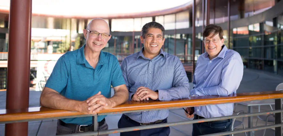
Photo by Aaron Kehoe: Device creators Scott L. Delp, Gabriel Sanchez, and Mark Schnitzer at the James H. Clark Center.
Stanford News - August 27th, 2024 - by Andrew Myers
In 2006, Stanford professors Scott Delp and Mark Schnitzer applied to the Stanford Bio-X Interdisciplinary Initiatives Program for a modest seed grant to explore a promising, but unproven, new medical imaging technique they hoped to develop. Their aim was to build upon a novel optical imaging technique, called microendoscopy, which Schnitzer had pioneered for viewing individual cells deep inside the tissues of live animals.
At the time, Delp, Schnitzer, and their students had gathered preliminary data showing that microendoscopy imaging in live mice could reveal sarcomeres, the force generating units of muscle. Thanks to the initial support from the Bio-X grant, and the efforts of Stanford PhD students Michael Llewellyn, Robert Barretto, Melinda Cromie (a Bio-X SIGF Fellow), and Gabriel Sanchez, the two laboratories developed microendoscopy into a technology that could be applied to visualize sarcomeres in awake and alert human subjects.
Seed grant symposium
For those interested in this and other transformative research, Bio-X will host the Stanford Bio-X Interdisciplinary Initiatives Seed Grants Program Symposium at the James H. Clark Center on Aug. 29 from 1:15 to 3:30 p.m. A poster session will follow.
After more than a decade in development, the fruits of that hard work paid off and propelled dermatological microendoscopy beyond research and toward the clinical realm. The result is a next-generation AI-powered imaging platform being taken to market by a company co-founded by the researchers to serve the broader health care field. The new company, Enspectra Health Inc., licenses the technology from the Stanford University Office of Technology Licensing.
True breakthrough

Photo by Aaron Kehoe: Device creators Scott L. Delp, Gabriel Sanchez, and Mark Schnitzer at the James H. Clark Center.
The company’s VIO Skin Platform was recently recognized with the FDA’s “Breakthrough Device” designation in select patient populations – a considerable accomplishment that expedites the FDA’s rigorous regulatory process. The platform non-invasively generates high-resolution digital images of live cells in living patients, allowing trained professionals to evaluate skin lesions in real time.
“The device is handheld, like an ultrasound apparatus. The entire imaging system can be carried in a little briefcase,” said Delp, who is a professor of bioengineering in the schools of Engineering and Medicine and an expert in medical technology development. “Our vision is that you put it against the skin and you get live-streamed images that will allow a doctor to identify if skin cancer is present.” No incisions. No long waits for biopsy results.
“It appears that the technology used in the new device represents the first new physical imaging modality to be FDA-cleared for clinical usage in about a quarter-century,” added Schnitzer, who is a professor of biology and applied physics in the School of Humanities and Sciences.
Two technologies in one
The device is intended to aid in the diagnosis of basal cell carcinoma and squamous cell carcinoma, which comprise most skin cancer cases in the United States. Skin cancer, however, is only the first application for this technology. Delp and Schnitzer believe it can be further developed to address many other clinical needs.
“Innovations in medical imaging have driven many of the key advances in modern medicine. We’re proud to continue that legacy into the cellular realm of living tissues,” said Sanchez.
The platform is not a single technology, but the unification of two approaches – one novel, the other mature. The newer approach, multiphoton optical imaging, allows the imaging of living cells without damage within the living body.
“It can visualize submicron structures,” said Delp. “You can image whole cells and subcellular structures in a living patient in the exam room.”
The more mature technology, reflectance confocal microscopy, around since the 1950s, allows the physician to see through the skin several layers deep to visualize skin features ranging from blood vessels, collagen, and pigment to the stratum corneum, hair follicles, hyperkeratosis, epidermal disarray, and more.
On to market
The combined imaging technologies will be complemented by AI-based predictive algorithms that allow trained dermatologists and pathologists to conduct non-invasive “virtual biopsies” in real time.
Only a select few new devices each year are considered for the FDA’s Breakthrough Device designation. All Breakthrough Devices must meet the FDA’s rigorous standards of safety and effectiveness. Working closely with Bio-X and faculty, the Office of Technology Licensing promotes the transfer of Stanford technology for society’s use and benefit while generating unrestricted income to support further research and education.
This story describes how Stanford research and discoveries are transferred for society’s use and benefit. It does not indicate an endorsement of any company.
originally published at Stanford News


