Stanford Report, September 30th, 2010
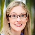
Bacteria have a way of sticking together. Lynette Cegelski, a Stanford assistant professor of chemistry, wants to unwind the secrets of exactly how they do it, the better to combat bacterial diseases.
Her research efforts just got a big boost – $1.5 million worth. She is one of three Stanford researchers, from chemistry, chemical engineering and the medical school, who have been chosen by the National Institutes of Health as winners of this year's New Innovator Awards.
Each of the faculty members will receive $1.5 million in research funds over five years. The award is designed to support unusually creative investigators at an early stage of their careers.
The NIH is also handing out a different flavor of research grants, known as the Transformative R01 Awards, to two other research projects, both in the medical school, that will explore exceptionally innovative, high-risk ideas.
New Innovator Awards
Cegelski uses new and unconventional approaches to understand the chemical and molecular basis of interactions in complex biological assemblies, such as the adhesive structures and aggregations formed by bacteria. Bacteria build fiber-like structures called curli to stick to inert surfaces, their hosts and each other. Adhering to one another enables bacteria to form biofilms, complex aggregations of bacteria that can play an important role in disease.
Cegelski and her group will study the architecture and function of curli and biofilms using chemical biology and solid-state nuclear magnetic resonance (NMR) spectroscopy, a method that can determine the structure and dynamics of interacting molecules at atomic resolution. Cegelski said she is inspired by the "suitability of solid-state NMR spectroscopy – particularly 'whole-cell NMR' – to deliver new clues to understanding macromolecular assembly events." Cegelski and her group will also search for novel antibiotics by testing the ability of small molecules to inhibit curli and biofilm assembly. "The NIH Director's New Innovator Award provides us with tremendous opportunity and freedom to tackle these more risky, but exciting, questions," Cegelski said.
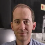
Alexander Dunn, an assistant professor of chemical engineering, studies molecular protein motors that generate mechanical force inside living cells. When functioning properly, mechanical forces help guide the changes in size and shape that occur during normal development of the embryo. When acting out-of-line, the forces generated by these tiny motors contribute to a cancer cell's ability to travel throughout the body.
"Cells communicate in part by pushing and tugging on their neighbors," Dunn said. Cells must cooperate with one another to develop into complicated, multicellular organisms. But cancer cells, rather than cooperate, ignore signals to stop dividing or to stay in one place.
Dunn and his group are using new light microscopy techniques to measure the forces generated by individual motor proteins in living cells to see how, when and where cells exert force on each other. Understanding these processes requires a research team with skills spanning genetics, cell biology, engineering and physics. "This grant makes it possible to put all of that talent together in one place," Dunn said.
Brian Feldman, an assistant professor of pediatric endocrinology in the School of Medicine, will use his $1.5 million award to study how hormones influence the fate of maturing stem cells.
His research on the basic biology of this relationship could lead to new therapies for a variety of diseases. In the long term, Feldman said, "We're trying to design new tools that will allow us to regulate stem cells in vivo. It might be possible to redirect stem cells towards more healthful lineages, or even induce their regenerative properties, without taking them out of a person." He imagines possibly heading off obesity by diverting stem cells away from the pathway that leads to fat cells, or treating muscle-wasting conditions by cueing stem cells to form more muscle.
Feldman will use the New Innovator Award to develop experimental systems, such as transgenic mice, to examine the regulation of stem cell fate in living animals. His prior research has shown that the circadian clock, the body's mechanism for setting daily rhythms, is embedded in specific hormone signaling pathways that regulate stem cell fate decisions. Feldman's lab is now creating transgenic mice with altered circadian clock function in their mesenchymal stem cells, the cells that give rise to tissues such as muscle, bone, cartilage and fat. The altered mesenchymal stem cells will be specially tagged with tracers to let the researchers follow them and reveal the potential of this approach to redirect stem cells' fates in a living animal.
Transformative R01 Awards
These awards will support projects that have the potential to create or overturn fundamental paradigms in biomedical research. The funding amount varies by project; in some instances, the funding amount has not yet been finalized.
Gary Peltz, a professor of anesthesia – in collaboration with research associate Toshi Nishimura and postdoctoral scholarYajing Hu – will use his $2.5 million over the next five years to study the effects of common drugs, including cholesterol medications, on mice with livers grown from human liver cells. One of the liver's main functions in the body is to break down, or metabolize, toxic chemicals, which can include drugs.
"What we will do is determine if the rate of metabolism of drugs that we test is determined by the genetic factors within the donor human liver cells that are used to reconstitute the human liver," Peltz said.
He expects that some livers will process a given drug more quickly than others, which would potentially change the drug's effects on the body. This study could provide insight into why people's responses to the same drug sometimes vary.
Marius Wernig, an assistant professor of pathology, and Thomas Sudhof, a professor of molecular and cellular physiology, discovered earlier this year that skin cells taken from mice can be converted directly into fully functional neuronal cells, skipping the intermediary stem cell step. Wernig and Sudhof will use their award funding (the exact amount is still being determined) over five years to see whether the same technique can be used on skin cells from humans.
Neurons grown from cells of patients with brain disease would preserve the genetic information contained in the original cell, enabling scientists to study the cellular mechanisms of disease up close in a way that imaging studies and postmortem analyses do not allow. This study will focus on neuropsychiatric diseases thought to arise from problems at the synapse – the space between neurons that signals must traverse to move from one cell to the next. But the potential is enormous. "It could be applicable to any brain disease you can think of," Wernig said of the new method.