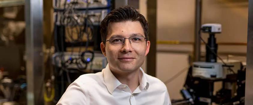
Photo by Steve Fisch: Sergiu Pasca, who coined the term "assembloids."
Stanford Medicine Scope - October 1st, 2021 - by Bruce Goldman
A recent article in the journal Nature credits Stanford Bio-X faculty and physician-neuroscientist Sergiu Pasca, MD, with blazing a trail toward a more profound understanding of early brain development, and of what can go wrong in the process, using a cell-based research innovation he named "assembloids."
In 2015, Pasca and his colleagues published a paper in Nature Methods describing a fascinating feat: His tinkering with induced pluripotent stem cells, or iPS cells -- former skin cells transformed so that they've acquired an almost magical capacity to generate all the tissues in the body -- had borne a three-dimensional product. From these "magic" iPS cells grew a complex conglomerate of cells capable of modeling specific organs.
Pasca's particular interest was in the brain, and in the experiments detailed in the study, his lab had caused human iPS cells to multiply and differentiate into small spherical clusters of brain tissue suspended in laboratory glassware.
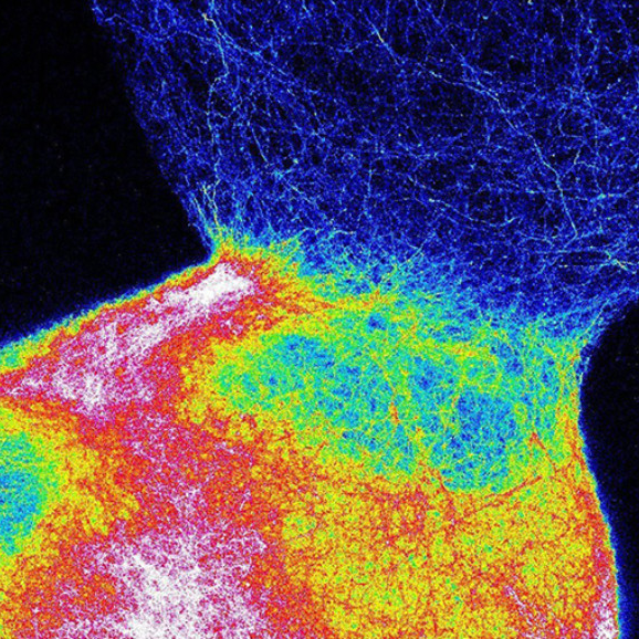
These clusters recapitulated the architecture and physiology of the human cerebral cortex -- the outermost layer of brain tissue, critical to perception, cognition and action. Pasca named these clusters, which grew to several millimeters in diameter and contained millions of cells, "cortical spheroids." Today, researchers around the world are using similar methodology to create models, broadly known as "organoids," to study other parts of the human body.
Two years later, in a study published in Nature, Pasca upped the ante by, first, generating a second kind of neural spheroid -- this time, representative of a deeper part of the developing forebrain called the subpallium -- and, second, by growing this kind of spheroid in conjunction with cortical spheroids, in the same dish.
To the researchers' amazement, spheroids of both types fused together, with nerve cells from subpallial spheroids migrating and poking extensions into the cortical spheroids and establishing working connections with nerve cells of a different type in the latter spheroids, just as occurs in fetal development.
"It's amazing that these cells already self-organize and know what they need to do," Pasca marveled in "Brain Balls," an article I wrote for our magazine, Stanford Medicine, a few years ago.
Pasca sensibly dubbed the two-fused-spheroid combos "assembloids," the Nature recap notes.
But why stop at two? Pasca has since created three-element assembloids composed of spheroids representative of cerebral cortex, spinal cord and skeletal muscle in order to model the circuitry of voluntary movement. He's also shown that stimulating the "cerebral cortex" spheroid can result in contraction of the "muscle" spheroid. (This accomplishment was published in Cell in late 2020.) He has explored other assembloid combinations, as well, such as the fusing of cortical spheroids with spheroids representing the striatum, a brain structure implicated in regulating our movements and responses to rewarding and aversive stimuli.
Because each spheroid begins with skin cells, they can be grown on a personalized basis -- and can therefore be extracted from patients with neurological disorders known or suspected to spring from early developmental aberrations (such as autism or schizophrenia). The cells can then be used to create models to probe these disorders' molecular, cellular and circuit-based deviations from the pathways of normal brain development, allowing scientists to study the brain in way they could never do with a living patient.
From the Nature article:
Assembloids are now at the leading edge of stem-cell research. Scientists are using them to investigate early events in organ development as tools for studying not only psychiatric disorders, but other types of disease as well.
An assembloid is by no means a complete, working brain. But, the article notes, "Pasca stands by the aphorism that all models are wrong, and some are useful. 'There's been important progress in the field in a short period of time,' he says."


