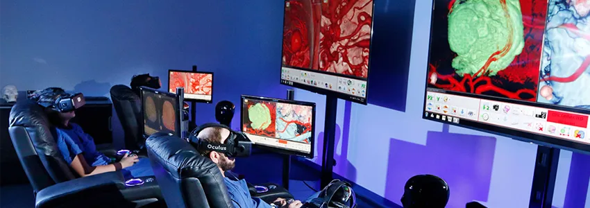
Photo by Paul Sakuma: The virtual reality system is helping train residents, assist surgeons in planning upcoming operations and educate patients. It also helps surgeons in the operating room, guiding them in a three-dimensional space.
Stanford Medicine News Center - July 11th, 2017 - by Mandy Erickson
Having undergone two aneurysm surgeries, Sandi Rodoni thought she understood everything about the procedure. But when it came time for her third surgery, the Watsonville, California, resident was treated to a virtual reality trip inside her own brain.
Stanford Medicine is using a new software system that combines imaging from MRIs, CT scans and angiograms to create a three-dimensional model that physicians and patients can see and manipulate — just like a virtual reality game.
After donning a headset connected to the VR system, Rodoni could clearly see the ballooning blood vessel, as well as the spot where her neurosurgeon, Gary Steinberg, MD, PhD, would place a clip to repair it. “Because I had been through this before, I thought I knew it all until I saw this,” she said. “I felt better knowing it was so clear to the doctor.”
Created by the Colorado startup Surgical Theater, the VR system is helping train residents, assist surgeons in planning upcoming operations and educate patients. It also helps surgeons in the operating room, guiding them in a three-dimensional space.
For the residents, class is held in a room in the hospital basement. Under low lighting, and surrounded by three massive screens, the residents settle into reclining chairs complete with drink holders — all promising a comfortable ride inside the human skull.
Once the residents don headsets, an instructor — who shows up as an avatar in a white coat — can lead them inside the brain of a patient. The system allows instructors to highlight different components of the brain, such as arteries to show an aneurysm, bones to show skull deformities or tissue to show a tumor, while rotating the view to illustrate how a tumor or aneurysm looks from different angles. They can also progress, as avatars, through the steps for removing a tumor or fixing an aneurysm, starting outside the skull.
‘A window into the brain’
Surgeons make their way down to the Neurosurgical Simulation Lab to practice an upcoming operation. Because they’re practicing on images from the actual patient, rather than a generic brain, they can map out the surgery ahead of time. “It’s a window into the brain — and a window into the brain of the particular patient we’re going to operate on,” said Anand Veeravagu, MD, an assistant professor of neurosurgery and the head of the Stanford Neurosurgical Simulation Lab.
The three-dimensional aspect of the imagery eases surgeons’ planning and improves the accuracy of the surgery, with the aim of producing safer procedures. “We can plan out how we can approach a tumor and avoid critical areas like the motor cortex or the sensory areas,” said Steinberg, professor and chair of neurosurgery. “Before, we didn’t have the ability to reconstruct it in three dimensions; we’d have to do it in our minds. This way it’s a three-dimensional rendering.”
Steinberg noted that in Rodoni’s case, an artery was attached to the top of the aneurysm. “You couldn’t see it on conventional imaging,” he said. “Had I not known about it, it could have been a real disaster.”
To show patients what’s going on inside their skulls, Malie Collins, MS, senior program lead for the VR program, rolls a mobile unit, complete with headset, into an examination or hospital room. Being able to see the problem in three dimensions reassures them, she said, adding that it’s especially useful for young patients or those who don’t understand English well. She can also download the imagery onto a thumb drive and give it to the patient as a souvenir.
“Traditionally, doctors can show their patient a standard physical model of the brain or of the spine and say, ‘On this model, imagine your tumor is located here,’” she said. “But with VR, we are able to immerse patients in their own anatomy, so they can very clearly get a sense of what’s going on.”
Stanford Medicine doctors are using the VR technology for the brain and spinal cord because these organs are stable and lend themselves to imagery — unlike other body parts, which move with blood flow and breathing. Collins said the technology may soon be available for the rest of the body.
‘Much, much more detail’
Surgeons typically use video feeds while they are operating, but the new VR technology adds a three-dimensional view which they can superimpose on the real-time video. “It has much, much more detail,” said Steinberg, the Bernard and Ronni Lacroute-William Randolph Hearst Professor in Neurosurgery and Neurosciences. For Rodoni’s surgery, “I had the 3-D rendering of her anatomy and could match that up with the surgical microscopic view, something I can’t do with any other technology.”
Veeravagu said some patients have chosen Stanford over other nearby hospitals solely because of the VR technology. “This software really helps them understand what it is they are about to undergo,” he said. “Seeing it on the screen, in 3-D, really helps put a patient’s mind at ease.”
It certainly did for Rodoni. Knowing where her aneurysm lay, and how Steinberg would repair it, helped calm her as she faced her third brain surgery. “I knew that Dr. Steinberg would be able to see the same thing I saw, and he wasn’t going to run into any surprises,” she said. Rodoni’s surgery went smoothly and she was discharged from the hospital within two days, her aneurysm gone.
 of Neurology")