Welcome to the biweekly electronic newsletter from Stanford Bio-X for members of the Bio-X Corporate Forum. Please contact us if you would like to be added or removed from this distribution list, or if you have any questions about Stanford Bio-X or Stanford University.
Highlights
** On October 9, 2013, Bio-X celebrated the 10th Anniversary of the James H. Clark Center, the hub of Bio-X. Check out CLARK CENTER @ 10X on the SPLASH PAGE as well as the Bio-X Timeline over the last 15 years!!
** Check out the article by Stanford President John Hennessy in the Nov/Dec 2013 issue of the Stanford Magazine on Bio-X and the Clark Center, "A Cauldron of Innovation".
Seed Grants
** UPDATE: Bio-X has just recently closed its 7th RFP for the IIP Seed Grants, and has received 141 Letters of Intent!
 SEED GRANTS FOR SUCCESS - Stanford Bio-X Interdisciplinary Initiatives Program (IIP)
The Bio-X Interdisciplinary Initiatives Program represents a key Stanford Initiative to address challenges in human health. The IIP awards approximately $3 million every other year in the form of two-year grants averaging about $150,000 each. From its inception in 2000 through the fifth round in 2010, the program has provided critical early-stage funding to 114 different interdisciplinary projects, involving collaborations from over 300 faculty members, and creating over 450 teams from five different Stanford schools. From just the first 5 rounds, the IIP awards have resulted in a 10-fold-plus return on investment, as well as hundreds of publications, dozens of patents filed, and most importantly, the acceleration of scientific discovery and innovation.
In 2012, Stanford Bio-X selected 23 new seed grant projects as the winners of the 6th round. Please go here to view the list of awardees, along with the titles of their projects and the abstracts of the research. Competition was intense as the awardees were chosen from 118 Letters of Intent (LOIs). Selection criteria included innovation, high-reward, and interdisciplinary collaboration. (To view the 114 other IIP projects that have been funded from the first 5 rounds, please click here.) In addition, SANOFI has also funded 4 new Bio-X IIP Seed Grant projects from round 6!
We are cultivating and are highly successful in building meaningful collaborations with numerous corporate colleagues. New collaborations through our seed grant projects are highly encouraged. To learn about how to get involved, please contact Dr. Hanwei Li or Dr. Heideh Fattaey.
IIP Seed Grants-Related Events
** On Monday, March 3, 2014, Bio-X had a Poster Session, featuring 105 different posters from research by all scientists within the Stanford Bio-X community. Over 250 people attended the session, which allowed for an excellent venue to discuss science and research with colleagues from both academia and industry.
** On Monday, August 26, 2013, Bio-X had its second annual IIP Symposium of the year at the Clark Center, which highlights projects that exemplify the Stanford Bio-X mission of crossing boundaries to bring about interdisciplinary research and solutions in the field of life bioscience. The symposium was a huge success with over 300 people attending this event, which included 8 oral presentations and 136 poster presentations. Recorded talks from the symposium will be uploaded soon. If you'd like to view the talks for previous symposia through the years, please click here.
Fellowships
Every year, graduate students and postdoctoral scholars of Bio-X affiliated faculty are highly encouraged to apply for the Bio-X Fellowships, which are awarded to research projects that are interdisciplinary and utilize the technologies of different fields to solve different biological questions. Students are encouraged to work collaboratively with professors of different departments, thus creating cross-disciplinary relationships among the different Stanford schools. Our fellows have conducted exciting research, resulting in publications in high-impact journals and have been offered excellent positions in industry and academia. To date, Stanford Bio-X has a total of 152 Fellows.
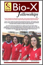
On June 26, 2013, Bio-X held its annual Bio-X Fellows Symposium, where there were four 15-minute oral presentations followed by one-minute spiels from current fellows. The 25 newest fellows selected this year were also announced, and about 100 attendees came to the symposium. Please click on the "Bio-X Fellows Symposium" link above for the agenda and titles of the talks, and on the icon of the brochure above for the updated and latest Bio-X Fellowships brochure.
To view the numerous projects that have been awarded over the years, please click here.
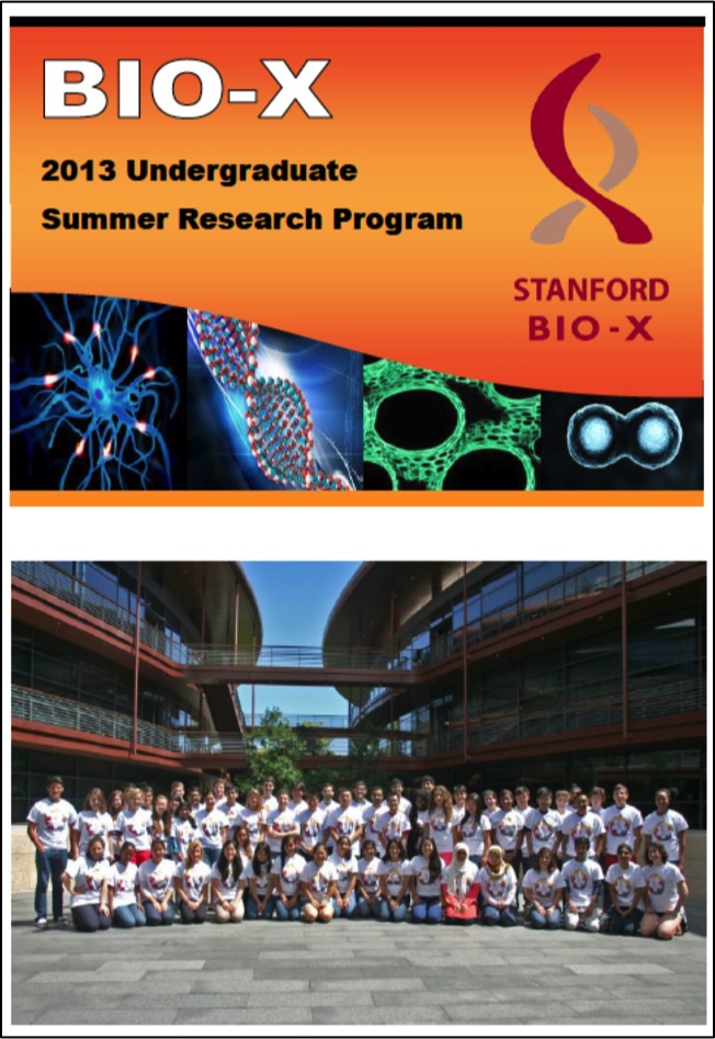 BIO-X UNDERGRADUATE SUMMER RESEARCH PROGRAM
The Bio-X Undergraduate Summer Research Program supports undergraduate research training through an award designed to support interdisciplinary undergraduate summer research projects. The program is an invaluable opportunity for students to conduct hands-on research, learn how to carry out experiments in the laboratory, and develop the skills to read and analyze scientific literature.
This program is eligible to Stanford students who want to work in the labs of Bio-X affiliated faculty. To date, 241 students have been awarded the opportunity to participate in the Bio-X Undergraduate Summer Research Program. This summer is Stanford Bio-X's 8th round of USRP.
Participating undergraduates are also required to present poster presentations on the research that they've conducted during the program. Please click here for title lists of past posters that our undergraduates have presented.
Many fruitful collaborations and relationships have been established with industry through fellowships. Please contact Dr. Hanwei Li or Dr. Heideh Fattaey if you'd like to learn more about how to get involved with these fellowship programs.
News
 "New Treatments for Blindness"
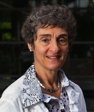
Bio-X Director Carla Shatz featured on broadcast
An episode on the Charlie Rose Brain Series, titled "New Treatments for Blindness" was broadcast on Channel Thirteen: New York Public Media and on PBS stations nationwide on Monday, April 21, 2014. Participants on this episode included: Charlie Rose, Eric Kandel (Columbia University), Carla Shatz (Stanford University), Jean Bennett (University of Pennsylvania), Steven Schwartz (Jules Stein Eye Institute, UCLA), Eberhart Zrenner (University of Tuebingen), and Sanford Greenberg (Johns Hopkins Wilmer Eye Institute). Please go here to view the broadcast.

 Stanford scientists observe brain activity in real time
Bio-X Affiliated Faculty Michael Lin and Mark Schnitzer
Bio-X IIP Seed Grant Project
Bio-X NeuroVentures Project
Two Stanford scientists have worked together to create tools for observing nerves in living animals that signal between themselves in real time. Observing the glowing trails of light spreading between connected nerves will help scientists understand how those individual signals add up to the complex collection of a person's thoughts and memories. "You want to know which neurons are firing, how they link together and how they represent information," said Michael Lin, assistant professor of pediatrics and of bioengineering. "A good probe to do that has been on the wish list for decades." Lin and Mark Schnitzer, associate professor of biology and of applied physics, developed two different approaches to allow neuroscientists to read brain activity more quickly and sensitively. Their research papers on this topic were published April 22 in Nature Neuroscience (Lin's study) and Nature Communications (Schnitzer's study).
 Stanford team makes switching off cells with light as easy as switching them on
Bio-X Affiliated Faculty Karl Deisseroth
In 2005, a Stanford University scientist discovered how to switch brain cells on or off with light pulses by using special proteins from microbes to pass electrical current into neurons. Since then, research teams around the world have used the technique that this scientist, Karl Deisseroth, MD, PhD, dubbed “optogenetics” to study not just brain cells but heart cells, stem cells and the vast array of cell types across biology that can be regulated by electrical signals — the movement of ions across cell membranes. Optogenetics gave researchers a powerful investigational technique to deepen their understanding of biological system design and function in animal models. But first-generation optogenetics had a shortcoming: Its light-sensitive proteins were potent at switching cells on, but less effective at turning them off. Now in a paper culminating years of effort, Deisseroth’s team has re-engineered their light-sensitive proteins to switch cells off far more efficiently than before. The paper was published April 25 in Science. “This is something we and others in the field have sought for a very long time,” said Deisseroth, senior author of the paper and professor of bioengineering and of psychiatry and behavioral sciences. Thomas Insel, MD, director of the National Institute of Mental Health, which funded the study, said this improved “off” switch will help researchers to better understand the brain circuits involved in behavior, thinking and emotion. “This latest discovery by the Deisseroth team is the kind of neurotechnology envisioned by President Obama when he launched the BRAIN Initiative a year ago,” Insel said. “It creates a powerful tool that allows neuroscientists to apply a brake in any specific circuit with millisecond precision, beyond the power of any existing technology.”
![]() Stanford bioengineers create circuit board modeled on the human brain
Stanford bioengineers create circuit board modeled on the human brain
Bio-X Affiliated Faculty Kwabena Boahen
Stanford bioengineers have developed a new circuit board modeled on the human brain, possibly opening up new frontiers in robotics and computing. For all their sophistication, computers pale in comparison to the brain. The modest cortex of the mouse, for instance, operates 9,000 times faster than a personal computer simulation of its functions. Not only is the PC slower, it takes 40,000 times more power to run, writes Kwabena Boahen, associate professor of bioengineering at Stanford, in an article for the Proceedings of the IEEE. "From a pure energy perspective, the brain is hard to match," says Boahen, whose article surveys how "neuromorphic" researchers in the United States and Europe are using silicon and software to build electronic systems that mimic neurons and synapses. Boahen and his team have developed Neurogrid, a circuit board consisting of 16 custom-designed "Neurocore" chips. Together these 16 chips can simulate 1 million neurons and billions of synaptic connections. The team designed these chips with power efficiency in mind. Their strategy was to enable certain synapses to share hardware circuits. The result was Neurogrid – a device about the size of an iPad that can simulate orders of magnitude more neurons and synapses than other brain mimics on the power it takes to run a tablet computer.
 Scientists develop 'playbook' for reverse engineering tissue
Bio-X Affiliated Faculty Steve Quake
Consider the marvel of the embryo. It begins as a glob of identical cells that change shape and function as they multiply to become the cells of our lungs, muscles, nerves and all the other specialized tissues of the body. Now, in a feat of reverse tissue engineering, Stanford University researchers have begun to unravel the complex genetic coding that allows embryonic cells to proliferate and transform into all of the specialized cells that perform myriad biological tasks. A team of interdisciplinary researchers took lung cells from the embryos of mice, choosing samples at different points in the development cycle. Using the new technique of single-cell genomic analysis, they recorded what genes were active in each cell at each point. Though they studied lung cells, their technique is applicable to any type of cell. "This lays out a playbook for how to do reverse tissue engineering," said Stephen Quake, PhD, the Lee Otterson Professor in the School of Engineering and a Howard Hughes Medical Institute investigator. The researchers' findings are described in a paper published online April 13 in Nature. Quake, who also is a professor of bioengineering and of applied physics, is the senior author. The lead authors are postdoctoral scholars Barbara Treutlein, PhD, and Doug Brownfield, PhD.
Mice with human livers would have saved lives if used in toxicology testing, study shows
Bio-X Affiliated Faculty Gary Peltz
Seven of 15 patients developed severe liver toxicity and died after taking a hepatitis B drug as part of a clinical trial in the early 1990s. Had a special variety of laboratory mouse been available then, that outcome could have been avoided, according to a study by researchers at the Stanford University School of Medicine. A few years ago, Gary Peltz, MD, PhD, professor of anesthesia, in collaboration with researchers at the Central Institute for Experimental Animals in Kawasaki, Japan, developed a line of bioengineered mice whose livers were largely replaced with human liver cells that recapitulate the architecture and function of a human liver. Published experiments have shown that using these bioengineered mice enables researchers to detect drugs' toxic metabolites and interactions with other drugs that do not occur in mouse livers. These would go unnoticed until human testing, and could produce severe adverse consequences in humans. Now, Peltz and his fellow researchers have demonstrated that the drug administered in that tragic trial two decades ago — which an investigation conducted by the National Academy of Sciences determined had shown no signs of being dangerous during rigorous preclinical toxicology tests — causes severe liver toxicity when given to the bioengineered mice with humanized livers. This observation would have served as a bright red stop signal that would have prevented the drug from being administered to humans. The researchers' findings were published online April 15 in PLoS Medicine. Peltz is senior author of the paper. Postdoctoral scholar Dan Xu, PhD, is lead author.
 Stanford research shows benefits of crystallization
Bio-X Affiliated Faculty Zhenan Bao
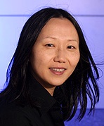
Sometimes engineers invent something before they fully comprehend why it works. To understand the "why," they must often create new tools and techniques in a virtuous cycle that improves the original invention while also advancing basic scientific knowledge. Such was the case about two years ago, when Stanford engineers discovered how to make a more efficient type of "strained organic semiconductors" that carry currents faster – a big step toward producing flexible electronic devices that couldn't be built using rigid silicon chips. Stanford chemical engineering Professor Zhenan Bao and her team discovered how to control the process through which those organic molecules assembled and crystallized as the liquid evaporated. Their findings are described in a recent Nature Communications study. Bao and her team wanted to understand why their process created such an electronically useful crystal lattice. So they launched a new experiment with help from organic thin film characterization expert Aram Amassian, an assistant professor at King Abdullah University of Science and Technology, or KAUST, in Saudi Arabia. The process of crystallization normally occurs in the blink of an eye and the researchers needed to understand it at the nanoscale. To do this they had to create a way to record and visualize molecules as they formed crystals in slow motion. In the report, Bao and Amassian reveal how they accomplished this feat: They combined a tiny, bright X-ray beam produced by the Cornell High Energy Synchrotron Source, or CHESS, in Ithaca, N.Y., with high-speed X-ray cameras to shoot a movie showing how organic molecules form different types of ordered structures or crystals.
Gene panel effectively screens dozens of genes for cancer-associated mutations, study finds
Bio-X Affiliated Faculty James Ford
As many as 10 percent of women with a personal or family history of breast or ovarian cancer have at least one genetic mutation that, if known, would prompt their doctors to recommend changes in their care, according to a new study by researchers at the Stanford University School of Medicine. The women in the study did not have mutations in BRCA1 or BRCA2 (mutations in these genes are strongly associated with hereditary breast and ovarian cancer), but they did have mutations in other cancer-associated genes. The study was conducted using what’s known as a multiple-gene panel to quickly and cheaply sequence just a few possible genetic culprits selected by researchers based on what is known about a disease. Although such panels are becoming widely clinically available, it’s not been clear whether their use can help patients or affect medical recommendations. “Although whole-genome sequencing can clearly be useful under the right conditions, it may be premature to consider doing on everyone,” said James Ford, MD, who directs Stanford’s Clinical Cancer Genetics Program. “Gene panels offer a middle ground between sequencing just a single gene like BRCA1 that we are certain is involved in disease risk, and sequencing every gene in the genome. It’s a focused approach that should allow us to capture the most relevant information.” Ford, an associate professor of medicine and of genetics, is the senior author of the study, published April 14 in the Journal of Clinical Oncology. Allison Kurian, MD, assistant professor of medicine and of health research and policy, and associate director of the Clinical Cancer Genetics Program, is the study’s lead author.
Gene variant puts women at higher risk of Alzheimer’s than it does men, study finds
Bio-X Affiliated Faculty Michael Greicius
Carrying a copy of a gene variant called ApoE4 confers a substantially greater risk for Alzheimer’s disease on women than it does on men, according to a new study by researchers at the Stanford University School of Medicine. The scientists arrived at their findings by analyzing data on large numbers of older individuals who were tracked over time and noting whether they had progressed from good health to mild cognitive impairment — from which most move on to develop Alzheimer’s disease within a few years — or to Alzheimer’s disease itself. The discovery holds implications for genetic counselors, clinicians and individual patients, as well as for clinical-trial designers. It could also help shed light on the underlying causes of Alzheimer’s disease, a progressive neurological syndrome that robs its victims of their memory and ability to reason. Its incidence increases exponentially after age 65. An estimated one in every eight people past that age in the United States has Alzheimer’s. Experts project that by mid-century, the number of Americans with Alzheimer’s will more than double from the current estimate of 5-6 million. According to the Alzheimer’s Association, it is already the nation’s most expensive disease, costing more than $200 billion annually. (The epidemiology of mild cognitive impairment is fuzzier, but this gateway syndrome is clearly more widespread than Alzheimer’s.)
 Scientists identify source of most cases of invasive bladder cancer
Bio-X Affiliated Faculty Philip Beachy
A single type of cell in the lining of the bladder is responsible for most cases of invasive bladder cancer, according to researchers at the Stanford University School of Medicine. Their study, conducted in mice, is the first to pinpoint the normal cell type that can give rise to invasive bladder cancers. It’s also the first to show that most bladder cancers and their associated precancerous lesions arise from just one cell, and explains why many human bladder cancers recur after therapy. “We’ve learned that, at an intermediate stage during cancer progression, a single cancer stem cell and its progeny can quickly and completely replace the entire bladder lining,” said Philip Beachy, PhD, professor of biochemistry and of developmental biology. “All of these cells have already taken several steps along the path to becoming an aggressive tumor. Thus, even when invasive carcinomas are successfully removed through surgery, this corrupted lining remains in place and has a high probability of progression.” Although the cancer stem cells, and the precancerous lesions they form in the bladder lining, universally express an important signaling protein called sonic hedgehog, the cells of subsequent invasive cancers invariably do not — a critical switch that appears vital for invasion and metastasis. This switch may explain certain confusing aspects of previous studies on the cellular origins of bladder cancer in humans. It also pinpoints a possible weak link in cancer progression that could be targeted by therapies.
Events
| Bio-X May 1, 2014, 3:15 pm - 4:15 pm Clark Center Auditorium, Stanford, CA Bio-X Frontiers in Interdisciplinary Biosciences Seminars - "Rare somatic cells from human breast tissue exhibit extensive lineage plasticity" Speaker: Thea D. Tlsty, UCSF |
Neurosciences Institute May 1, 2014, 2014, 12 pm - 1 pm Clark Center Auditorium, Stanford, CA "Cross-model cortical plasticity" Speaker: Hey-Kyoung Lee, PhD, Mind/Brain Inst |
| Cardiovascular Institute May 6, 2014, 2014, 12 pm - 1 pm LKSC 120, Stanford, CA Frontiers in Cardiovascular Science - "Cardio-oncology: New insights from MRI regarding cancer treatment related cardiovascular injury" Speaker: Gregory Hundley, MD |
Microbiology & Immunology May 7, 2014, 12 pm - 1 pm Munzer Auditorium, Stanford, CA "A global view of TB pathogenesis" Speaker: Chris Sassetti, U Mass |
Resources
| Stanford University |
| Stanford Bio-X |
| Bio-X Seed Grants The Stanford Bio-X Interdisciplinary Initiatives Program (IIP) provides seed funding for high-risk, high-reward, collaborative projects across the university, and have been highly successful in fostering transformative research. |
| Office of Technology and Licensing "Techfinder" Search the OTL Technology Portal to find technologies available for licensing from Stanford. |
| Stanford Center for Professional Development - Take advantage of your FREE membership! - Take online graduate courses in engineering, leadership and management, bioscience, and more. - Register for free webinars and seminars, and gets discounts on courses. |
| Stanford Biodesign Video Tutorials on how FDA approves medical devices A series of video briefs recently produced by the Stanford Biodesign Program teaches innovators how to get a medical device approved for use in the United States. This free, online library of 60 videos provides detailed information on the Food and Drug Administration regulatory process, short case studies and advice on interacting with the FDA. |
To learn more about Stanford Bio-X or Stanford University, please contact Dr. Hanwei Li, the Bio-X Corporate Forum Liaison, at 650-725-1523 or lhanwei1@stanford.edu, or Dr. Heideh Fattaey, the Executive Director of Bio-X Operations and Programs, at 650-799-1608 or hfattaey@stanford.edu.


