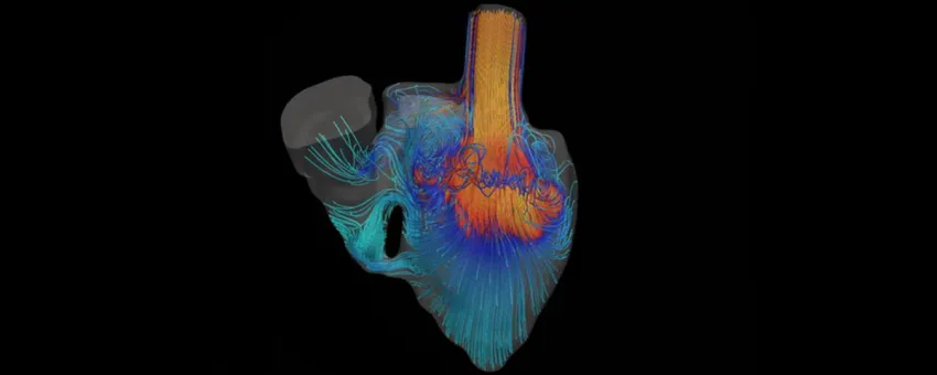
Image courtesy Marsden Lab: A simulation shows blood flowing through the heart of a baby born with a defect. The idea is to help in planning for surgery. Watch the video!
Stanford Engineering - January 21st, 2017
MRI and CT scans reveal what’s happening inside a patient’s body. Surgeons often study such scans when planning operations. Alison Marsden, an associate professor of bioengineering, wants to make such diagnostic imagery even more useful by creating patient-specific software simulations.
“When you come into the hospital and get scanned in the MRI machine or a CT scanner, what we often get is a beautiful picture of your anatomy, “ Marsden says. “But we don’t get often this detailed picture of how the blood is flowing, recirculating and moving through the blood vessels. And we also can’t use the imaging to make predictions.”
At Stanford’s Cardiovascular Biomechanics Computational Lab, Marsden and her colleagues are developing a new generation of software simulations to help plan surgeries. “What we’re trying to do is bring in that missing piece of what are these detailed blood flow patterns and what might happen if we go in and make an intervention,” she says, adding, “for example, opening up a blocked blood vessel or putting in a bypass graft.”
 and of Bioengineering and (by courtesy) of Mechanical Engineering")