Photo by ancroft, Shutterstock.
Stanford Medicine Scope - July 25th, 2017 - by Tracie White
In March, I wrote a blog post about a Stanford study that showed moderate exercise may be good for patients with hypertrophic cardiomyopathy, the heart disease most commonly associated with sudden death in athletes. This contradicted the general medical consensus that people with HCM should be extremely cautious about exercising and should not participate in any competitive sports.
Now, two new Stanford studies explore the uses of treadmill exercise testing along with sonograms of the heart as an improved method of determining treatment plans for patients with two types of cardiomyopathy, hypertrophic and dilated.
“The ultrasound enables us to view how the heart contracts while the exercise stress test helps us to look at how the body is using oxygen,” said Kegan Moneghetti, MD, a postdoctoral scholar at Stanford and first author of both studies. “By combining the two, you get incremental prognostic power.”
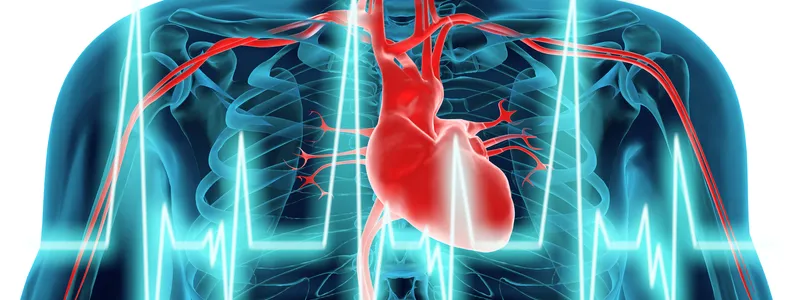
Cardiomyopathies are a group of serious disorders of the heart muscle with different causes. Hypertrophic cardiomyopathy is a genetic disorder that causes a thickening of the heart muscle and therefore leads to problems with blood flow. Dilated cardiomyopathy, which has a variety of causes from ranging from high blood pressure to ischemic heart disease, results in a weakened and enlarged heart that doesn’t pump enough blood to the rest of the body.
The first of the two studies, published online in the American Journal of Cardiology, examined 131 patients who had previously undergone the combined heart sonogram and exercise testing. The patients were followed for more than four years. The testing results were compared with 80 patients who didn’t undergo the combined procedure.
In the second study, published in the European Heart Journal – Cardiovascular Imaging, researchers looked at 208 patients with dilated cardiomyopathy who underwent the same procedure.
Both studies showed the combined exercise testing provided an improved model for routine examination of these cardiomyopathy diseases that could possibly reduce future risks of the disease. The tests were used to measure risks of death from the disease and whether the patient might need a ventricular assist device to help the heart pump or a heart transplant.
“Exercise has always been promoted as being great for health,” Moneghetti said. “We wanted to show how it can be better incorporated in diagnostics in cardiomyopathy patients.”
The studies examined the use of this test just once after the patients’ were diagnosed. In practice, the patients usually have the tests done annually at Stanford.
The process involves conducting a heart sonogram both immediately before and then after the patient does a 10 to 15-minute exercise test on a treadmill. The patient runs on a treadmill while breathing into a mask that measures carbon dioxide and oxygen levels. A heart sonogram technician slides a microphone-like device called a transducer over the chest area. This allows reflected sound waves to provide a live picture of the heart and valves.
“We think this could provide a new approach to thinking about the assessment of cardiomyopathy,” Moneghetti told me.
The studies also provided evidence that in a controlled environment, with constant cardiovascular examination going on, exercise testing appears safe for these patients, Moneghetti said.