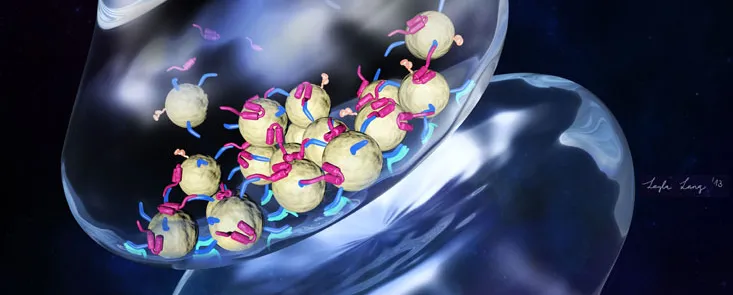
Graphic © 2013 by Layla Lang: The drawing shows two adjacent nerve cells, with the top cell containing neurotransmitters housed in bubble-like packets (yellow) that cluster within the cell's bulbous protrusions. Biochemical events cause these packets to fuse with the cell's outer membrane, spilling their contents into the tiny space separating it from the adjacent nerve cell. The packets' clustering is carefully regulated by several proteins. One end of the protein alpha-synuclein (magenta) binds to a different protein (blue) embedded in packets' surfaces, while the other end is attracted to the packets' surfaces themselves. The effect is to bring packets together.
Inside Stanford Medicine - April 30, 2013 - by Bruce Goldman
Researchers at the Stanford University School of Medicine have exposed the possible function, in the healthy brain, of a mysterious molecule that has been strongly implicated in Parkinson's disease, a degenerative disorder of the central nervous system. They made their discovery using a stripped-down experimental system that mimics key aspects of how nerve cells communicate with one another.
Thomas Südhof, MD, has devoted much of his career to understanding the goings-on in the terrifically complex nozzles from which nerve cells squirt specialized signaling chemicals called neurotransmitters. It is the diffusion of neurotransmitters from one nerve cell to the next that underpins our thoughts, feelings and movements.
The brain's activity is no mere mob-action squirt-gun melee, though. For our most exalted organ to do its job, the signals sent by nerve cells must be marked by profound precision, both in their intensity and in their timing. The details of how this occurs are still coming into focus.
"We'd just developed this new system, and we were scouting out interesting proteins implicated in nerve-cell signal transmission or, perhaps, in disease states," Brunger said.
Axel Brunger, PhD, devised a simplified experimental system that captured some of those inner workings, and was looking for interesting problems to solve with it. So he began sitting in on the Südhof lab's weekly meetings. "We'd just developed this new system, and we were scouting out interesting proteins implicated in nerve-cell signal transmission or, perhaps, in disease states," Brunger said.
Both Südhof and Brunger are professors of molecular and cellular physiology as well as Howard Hughes Medical Institute investigators. They share senior authorship of a paper, published April 30 in eLife, describing the advance that came from the ensuing collaboration.
Brunger's new experimental system intrigued Jacqueline Burré, PhD, a research associate in Südhof's group, who was subjecting a protein called alpha-synuclein to close scrutiny. Alpha-synuclein-rich clumps called Lewy bodies abound in the brains of Parkinson's patients and are considered a hallmark of the disease.
This would seem to implicate an overabundance of alpha-synuclein as a culprit in Parkinson's disease, but the truth appears to be more nuanced. Südhof's group, for example, had created a strain of lab mice lacking the gene for alpha-synuclein and also the genes for two close cousins of that protein, beta- and gamma-synuclein. Burré found that, curiously, these mice seemed to function just fine in the absence of any Greek-letter-prefaced synuclein — at first. The youthful "triple knockouts," as they are known, thrived in their early days, but the absence of synucleins seemed to catch up with them as they aged. The mice eventually developed serious deficiencies in motor function and died much sooner than their normal peers. (Parkinson's patients develop movement-related symptoms including tremor in the extremities and stiffness.) The effect was analogous to running a car for several years without changing the oil. Despite the lack of immediately visible damage, the long-run effect is almost certainly the engine's demise.
"Lots of people have been looking at what alpha-synuclein does wrong," said Burré, who shares lead authorship of the eLife study with biophysicist Jiajie Diao, PhD, a research specialist in Brunger's group. "Almost no one is looking at what it does normally, in a healthy person or animal. That was our prime focus, and we wanted to know how this works at the molecular level."
Brunger's experimental system, in which the rapid-fire molecular events involved in neurotransmitter secretion are pared down to the basics, looked like an excellent way to find out.
Nerve cells don't simply squirt out neurotransmitters willy-nilly. Within the complex networks that constitute our brains, every individual nerve cell has a lengthy, snaking, tubular extension cord, or axon, that hooks up with thousands of other nerve cells. Neurotransmitters are housed within tiny bubble-like packets in the cell. These packets congregate in myriad small, bulbous nozzles dotting the axon, with each bulb abutting a downstream nerve cell. When an electrical impulse travels down the axon on which those bulbs reside, it triggers the fusion of the neurotransmitter-packed packets with the nerve cell's outer membrane. The packets' contents then spill into the narrow space separating the bulbs from the nerve cells they abut.
Südhof's group has been instrumental in characterizing several proteins - some embedded in the surfaces of all neurotransmitter-loaded packets, and others in the membranes enclosing the cells' bulbous nozzles — that, in response to electrical impulses coursing through the nerve cell, shift their structures temporarily and, like a living zipper, interact in such a way as to pull the packets and the cell's outer membrane so tightly together that they fuse and the neurotransmitters are released.
Brunger's bench-top system employs two kinds of synthetic vesicles: very small ones to represent the tiny packets, and others representing the secretory bulbs. In their experiments, the Stanford investigators coated both types of vesicles, respectively, with proteins that are specifically found on the packet or the bulb surfaces and that have been shown to be essential to proper membrane fusion. Substituting a chemical procedure for the electrical impulses that normally elicit these proteins' interactions, the scientists could trigger fusion between the two vesicle types at will. Thus, the system served as a simplified synthetic model of the apparatus of nerve-cell secretion.
Next, the scientists measured how many of the packet vesicles adhered to the surface of the bulb vesicles in the presence or absence of alpha-synuclein. Interestingly, alpha-synuclein's presence didn't seem to change the dynamics of the membrane-fusion process itself. Instead, the protein seemed to tether packet vesicles together. "Alpha-synuclein caused the packet vesicles to cluster together at the synthetic 'cell surface' membrane," said Burré.
After showing that this clustering effect was proportional to the amount of alpha-synuclein added, the team determined that it occurs because one part of alpha-synuclein has an affinity for the fatty substances that are the major constituents of the neurotransmitter-carrying packets' surfaces, while another part of the molecule binds to a particular protein that, in the experiment as well as in actual nerve cells, was embedded in the surfaces of the packets.
This could explain how both a dearth of functional alpha-synuclein resulting from a mutation in the gene coding for it and an overabundance of alpha-synuclein caused by, among other things, duplication of the gene can cause trouble, Burré said. In the first case, the dysfunctional product of the gene fails to induce enough clustering, so that an insufficient number of neurotransmitter-carrying packets are in place (near the transmission bulbs' surfaces) when they're needed. In the second case, a hyper-abundance of alpha-synuclein leads to its aggregation, interfering with the correct positioning of the packet vesicles near the transmission bulbs' surfaces or with their efficient fusion with those surfaces.
"Whether this is exactly what is happening in the living brain remains to be seen," cautioned Brunger. "We're working with a minimal system. We can't be sure yet if this handful of proteins we're adding to our reconstituted membranes are enough to fully recapitulate the natural process." But both his and Südhof's groups are actively looking for ways to replicate their findings in brain tissue.
Assuming they do, it still won't necessarily translate into the immediate development of Parkinson's drugs, Burré noted, because ensuring that precisely the right alpha-synuclein levels are present in people's nerve cells at the right times is tougher than just reducing or increasing levels of this protein. But the scientists say that the more we can learn about alpha-synuclein's normal function, the better. "You should try to completely understand the normal function of a protein before playing around with it in human subjects," she said.
Other study co-authors were postdoctoral scholars Sandro Vivona, PhD, Daniel Cipriano, PhD, and Minjoung Kyoung, PhD; and research associate Manu Sharma, PhD.
The study was funded by the HHMI and by the National Institutes of Health (grants R37MH63105 and NS077906).
Information on the medical school's Department of Molecular and Cellular Physiology, which also supported this work, is available at http://mcp.stanford.edu/.


