Inside Stanford Medicine - November 21, 2011 - by Christopher Vaughn
Stanford researchers have melded tools and technologies from engineering, computer science and stem cell biology to analyze hundreds of individual cancer cells and draw the most accurate portrait yet of the cellular composition of human colon cancer tissues.
In doing so, they have shown that the development of cancer is a kind of caricature of normal tissue development, and have discovered markers that allow them to gauge more accurately how dangerous a cancer is likely to be. They hope the work will lead to better and more targeted cancer therapies.
Using these technologies to analyze the gene-expression patterns of single cells, one by one, allows the researchers to dissect their different identities and clarify the physiology of complex tissue systems.
In tumor tissues, not all cancer cells are created equal. Solid tumors usually contain many kinds of cancer cells, each of which might be more or less dangerous, and may vary in how they respond to various anti-cancer therapies. One major problem researchers have had in understanding cancer is that when they analyze a cancerous lesion, they usually look at whole-tumor samples that contain a mixture of all these cells types. Amidst this diversity, spotting the signs that betray the really dangerous cells is difficult, like trying to draw the psychological profile of a murderer by sending out questionnaires to everyone who ever committed a legal infraction.
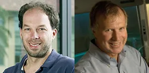
In a paper published online Nov. 13 in Nature Biotechnology, a team led by Stephen Quake, PhD, and Michael Clarke, MD describes how they used the single-cell PCR microfluidic technology invented in the Quake laboratory to analyze the individual gene-expression profile of hundreds of single colon cancer cells, which then they grouped into different subtypes.
“We have spent the last several years developing single-cell genomic measurement technologies,” said Quake, the Lee Otterson Professor in the School of Engineering and a professor of bioengineering and of applied physics. “To finally get all the tools together and produce a biologically important result is very gratifying.”
Using these technologies to analyze the gene-expression patterns of single cells, one by one, allows the researchers to dissect their different identities and clarify the physiology of complex tissue systems, explained Piero Dalerba, MD, instructor of medicine and co-first author of the study. The two other first authors are research associate Tomer Kalisky, PhD, and Debashis Sahoo, PhD, instructor of pathology. Taking advantage of a computer search technique newly developed by Sahoo, the authors identified 47 differentially expressed genes that are useful for the visualization of the various kinds of cells in the tumor.
“In a single experiment, we can now take a ‘snapshot’ of the cell composition of a specific tissue, visualize its different cellular subsets and easily discover novel markers to define them,” said Kalisky.
This methodology is important for the study of minority populations of cells, such as stem and progenitor cells, said Clarke, professor of oncology and the Karel H. and Avice N. Beekhuis Professor in Cancer Biology. “Using these techniques, we have cut a decade off the attempt to understand cellular hierarchy in tissues.”
The investigators noted that the study resulted from the close working relationships between researchers in various departments. “This project is the product of a fantastic synergy between engineers, computer scientists, stem cell biologists and medical oncologists, made possible by the interdisciplinary environment we have here at Stanford,” said Sahoo.
One of the key results of the work is that it revealed why there are many kinds of cancer cells in a tumor. One theory has been that cancer cells have an unstable genome and change in random ways as they reproduce. This research showed that colon cancer cells change along the same developmental lines followed by normal colon cells during differentiation processes. Like a caricature portrait, which is recognizably someone’s face even though certain features are exaggerated, the underlying order in cancer cell development is recognizably the same as in normal cells, although the percentage of individual cell types is abnormally expanded or reduced.
“What we see is that the cellular heterogeneity of colon-tumor tissues mirrors and recapitulates the lineage diversity of the normal colon epithelium, including both immature progenitor-like cells and more mature, specialized cells,” said Dalerba.
The researchers also showed that all the types of cancer cells in a colon tumor could spring from the differentiation of a single cancer cell, a process reminiscent of a stem-cell system.
One aspect of the research with potential clinical applications was the finding that tumors with a gene-expression profile similar to that of more immature cell types were more dangerous than those whose profile resembled more differentiated, specialized cells. The researchers developed a two-gene classification system that could be used to accurately predict how lethal a colon tumor was likely to be. They also showed that this simple, two-gene system predicted clinical outcomes more accurately than pathological grading of tumor samples, which is one of the few important parameters used by pathologists to define a tumor’s aggressiveness.
The researchers hope that use of this technology will lead to therapies that can target the most dangerous cells in a tumor.
Other Stanford co-authors were Michael Rothenberg, MD, clinical instructor of gastroenterology and hepatology; Anne Leyrat, PhD, a former visiting scientist; technicians Sopheak Sim and Jennifer Okamoto; graduate students Darius Johnston, Pradeep Rajendran and Jianbin Wang; senior research associate Dalong Qian, MD; tissue bank manager Janet Bueno; genomics core director Norma Neff, PhD; Andrew Shelton, MD, clinical associate professor of surgery; Brendan Visser, MD, assistant professor of surgery; postdoctoral scholars Maider Zabala, PhD, and Shigeo Hisamori, MD; and former postdoctoral scholar Yohei Shimono, MD.
This study was supported by grants from the National Institutes of Health and the Department of Defense. Additional support came from the California Institute for Regenerative Medicine, the Siebel Stem Cell Institute and the Thomas and Stacey Siebel Foundation, the Machiah Foundation and a BD Biosciences Stem Cell Research Grant.
Stanford’s departments of Bioengineering and of Medicine also supported the work. The Department of Bioengineering is jointly operated by the School of Engineering and the School of Medicine.


