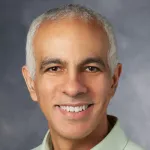
Dr. Michael Zeineh received a B.S. in Biology at Caltech in 1995 and obtained his M.D.-Ph.D. from UCLA in 2003. After internship also at UCLA, he went on to radiology residency and neuroradiology fellowship both at Stanford. He has been faculty in Stanford Neuroradiology since 2010. He spearheads many initiatives in advanced clinical imaging at Stanford, including clinical fMRI and DTI.
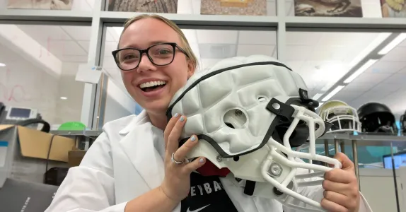
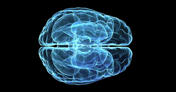
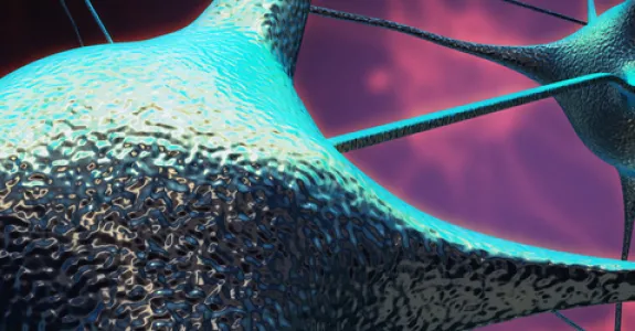
Dr. Michael Zeineh’s lab focuses on using imaging techniques to identify abnormalities in neurodegenerative disorders, particularly those affecting the microcircuitry in regions such as the hippocampal formation. Through in vivo, ex vivo (i.e. post-mortem), and culture methods, the lab has uncovered alterations in iron in the hippocampus that may be causative in Alzheimer’s Disease and underlie the inflammation that is synergistic with amyloid and tau. Recent longitudinal studies advanced MRI techniques to image athletes in high-impact sports have also revealed insights into the mechanisms behind mild traumatic brain injury, including concussion. The interdisciplinary lab has discovered changes that may underly chronic fatigue syndrome, which carries many similarities to Long COVID. The armamentarium of techniques employed includes ultra-high resolution 7.0T MRI & PET-MR (including DTI & QSM), advanced histology, X-ray microscopy and scattered light imaging (with Marios Georgiadis), electron microscopy, as well as exosomal analyses and Raman imaging (with Utkan Demirci). Emerging projects include animal models of mild traumatic brain injury (with David Camarillo), ultra-high field MRI applied to multiple neurological disorders (with Kawin Setsompop/Jon Polimeni), sodium imaging/DTI in epilepsy (with Fernando Boada/Jennifer McNab).