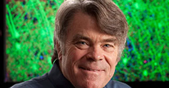
Now enjoying emeritus status, Prof. Stephen Smith remains active with work in computational neuroscience microscopy and genomic data science. His recent explorations have unearthed transcriptomic evidence for a previous unrecognized ubiquity of local neuropeptide signaling and possible involvement of same in memory engram formation. Smith led an active Stanford laboratory (1990-2014) that explored brain development, structure, function, and disease progression. The lab’s experimental approach typically began with invention of a new imaging method followed by applications of that method to attack previously intractable experimental challenges. Early on, Smith invented a novel fiber-optic spectrometer for calcium sensing that enabled the first detection and measurement of calcium transients in vertebrate neurons, the first quantitative measurements of presynaptic Ca transients, and the extraordinarily significant discovery of Ca influx through NMDA receptor channels. Later inventions led to numerous significant neuroscience discoveries, including retrograde actin flow within neuronal growth cones, intracellular Ca waves in astrocytes, the active role of dendritic filopodia in synaptogenesis, and the packeted delivery of synaptic protein components during synaptogenesis, and to the first optical measurements of single synaptic vesicle release, the first in vivo imaging of synaptotropic dendrite growth, and the first in vivo functional imaging measurements of visual receptive field development in a vertebrate animal. Smith’s laboratory also invented a unique and now widely used high-resolution proteomic imaging method called “Array Tomography” and applied the method to explore the molecular architecture of cortical microcircuits in mouse and human. Smith went emeritus and closed his Stanford laboratory in 2014, taking an exciting new position as Senior Investigator at the Allen Institute for Brain Science in his hometown of Seattle, Washington. At the Allen Institute he freshened up his data science proficiencies, driven by that Institutes prodigious production of extremely high-quality neuroscience data. He is now an Allen Institute Investigator Emeritus and an Allen Neural Dynamics Fellow and plans a return to California and Stanford campus life in Spring 2024.